Figure 1. Morphological observations. One of the corneoscleral rim used in the study. The black circle shows where the corneoscleral
rim was trimmed to separate the limbal explant from the whitish scleral tissue (A). Phase contrast microscopic shows the outgrowth of limbal stromal cells from the limbal explants on day 7 (magnification:
40×; B). Confluent culture of limbal stromal cells shows spindle morphology with petal growing pattern (magnification: 40×; C); Sphere formation by the limbal stromal cells when cultured with complete media without matrigel (D).
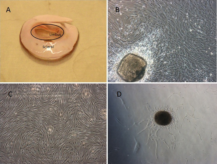
 Figure 1 of
Lim, Mol Vis 2012; 18:1289-1300.
Figure 1 of
Lim, Mol Vis 2012; 18:1289-1300.