Figure 5. Sphere culture of newborn mouse lacrimal gland epithelial cells. Spheres were clearly formed after 10 days (A). When spheres was dissociated with trypsin/EDTA and subcultured for 10 days, sphere formation was regenerated (B). When spheres were placed in tissue culture treated dishes for 4 days, the spheres attached to the dishes and cells were
expanded (C). When isolated spheres were cultured and embedded in collagen type I for 8 days, bundle-like cells expanded from the spheres
(D). Scale bars, (A) 100 µm; (B) 200 µm; (C, D) 400 µm.
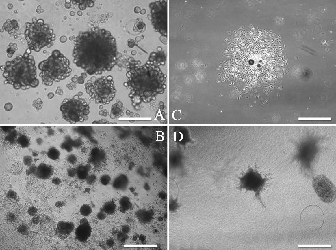
 Figure 5 of
Kobayashi, Mol Vis 2012; 18:1271-1277.
Figure 5 of
Kobayashi, Mol Vis 2012; 18:1271-1277.