Figure 3. PIM staining for the
hypoxic region. Eyes from different genotypes were stained with
antibodies against PIM. PIM signal (Green) was strong in Cited2CKO
compared to the control. Eyes collected from Cited2fl/fl;HIFfl/fl;Le-Cre+
showed a weaker signal compared to Cited2CKO
mice. Saline injection was used as a negative control.
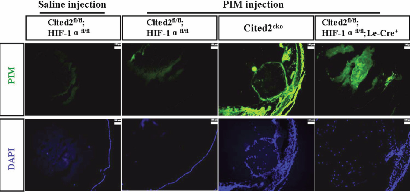
 Figure 3
of Huang, Mol Vis 2012; 18:1260-1270.
Figure 3
of Huang, Mol Vis 2012; 18:1260-1270.