Figure 2. Le-Cre mediated deletion of
Cited2 and HIF-1α and phenotypic rescue by HIF-1α
deletion. A: The expression levels of Cited2 and
HIF-1α were decreased in Cited2fl/fl;HIFfl/fl;Le-Cre+
mice compared to the control (n=3, *p<0.05). B:
Increased VEGF in Cited2CKO resumed
to the normal level after HIF-1α was deleted in Cited2CKO
mice. C: Under dissecting microscope, eyes collected
from Cited2fl/fl;HIFfl/fl;Le-Cre+
mice showed smaller sizes and cornea opacity (upper Panel).
H&E staining showed smaller lens and lens stalk formation at
the anterior part of the lens in Cited2fl/fl;HIFfl/+;Le-Cre+
and Cited2fl/fl;HIFfl/fl;Le-Cre+
mice. The insets represent higher magnification of the areas
indicated by arrows. D: Expression level of Cited2
(magenta) was low in Cited2fl/fl;HIFfl/fl;Le-Cre+
mice compared to the control . H&E pictures were taken at 5×
magnification to show the entire eye structure. DAPI pictures
were taken at 10× using adjacent sections. Red box indicates the
area shown in the counterstained pictures (20×). Scale bar in
each picture indicates 100 μm. E: α-SMA, F4/80, and CD31
protein expression indicated the cell types in the hyaloid
vascular system. Pictures were taken at 20× to show the
red-boxed area. Scale bar in each picture indicates 100 μm. A↔P:
anterior and posterior orientation of the eye.
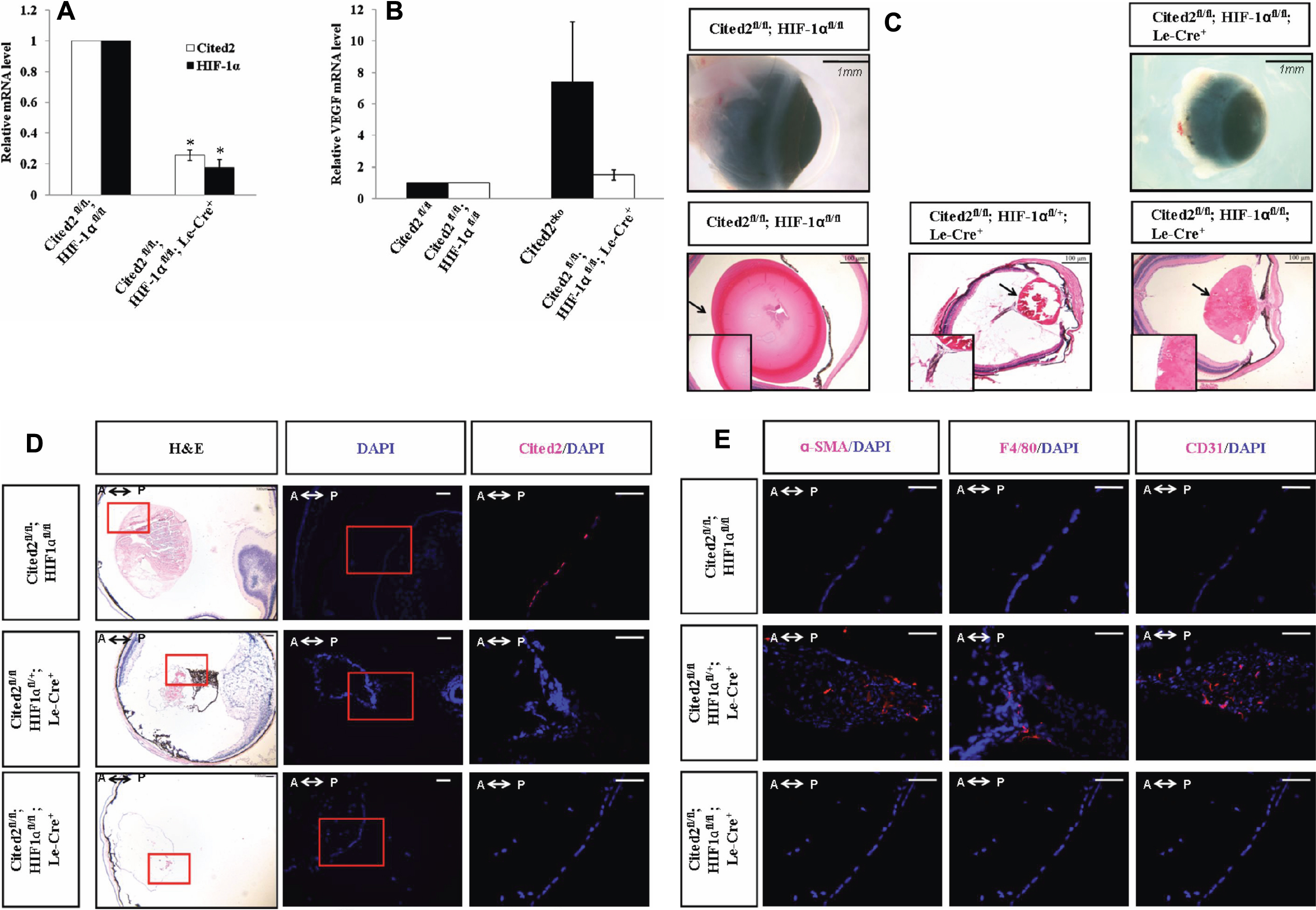
 Figure 2
of Huang, Mol Vis 2012; 18:1260-1270.
Figure 2
of Huang, Mol Vis 2012; 18:1260-1270.