Figure 1. Le-Cre mediated deletion of
Cited2 and morphological changes in Cited2CKO
mice. A: Expression of Cited2 was decreased in Cited2CKO
mice compared to control (n=3, *p<0.05). Modest increase of HIF-1α
and substantial increase of VEGF were observed in Cited2CKO
mice. B: Under a dissecting microscope, eyes collected
from Cited2CKO mice showed smaller sizes and
cornea opacity (upper panel). H&E staining showed smaller
lens, lens stalk (LS) formation at the anterior side of the
lens, and hyaloid hypercellularity and aberrant vasculature at
the posterior side of the lens in Cited2CKO
mice (lower panel). The insets represent higher magnification of
the areas indicated by arrows. C: Immunostaining with
Cited2 antibody showed decreased expression of Cited2 (magenta)
in Cited2CKO mouse lens. Counterstaining with
DAPI (blue) indicated that Cited2 localized in the nucleus.
H&E pictures were taken at 5× magnification to show the
entire eye structure. DAPI pictures were taken at 10× using
adjacent sections. Red box indicates the area shown in the
Cited2/DAPI counterstained pictures (20×). Scale bar in each
picture indicates 100 μm. D: Immunostaining with α-SMA,
F4/80, and CD31 (red) showed composition of the hyaloid vascular
system. Pictures were taken at 20× to show the red-boxed area.
Scale bar in each picture indicates 100 μm. A↔P: anterior and
posterior orientation of the eye.
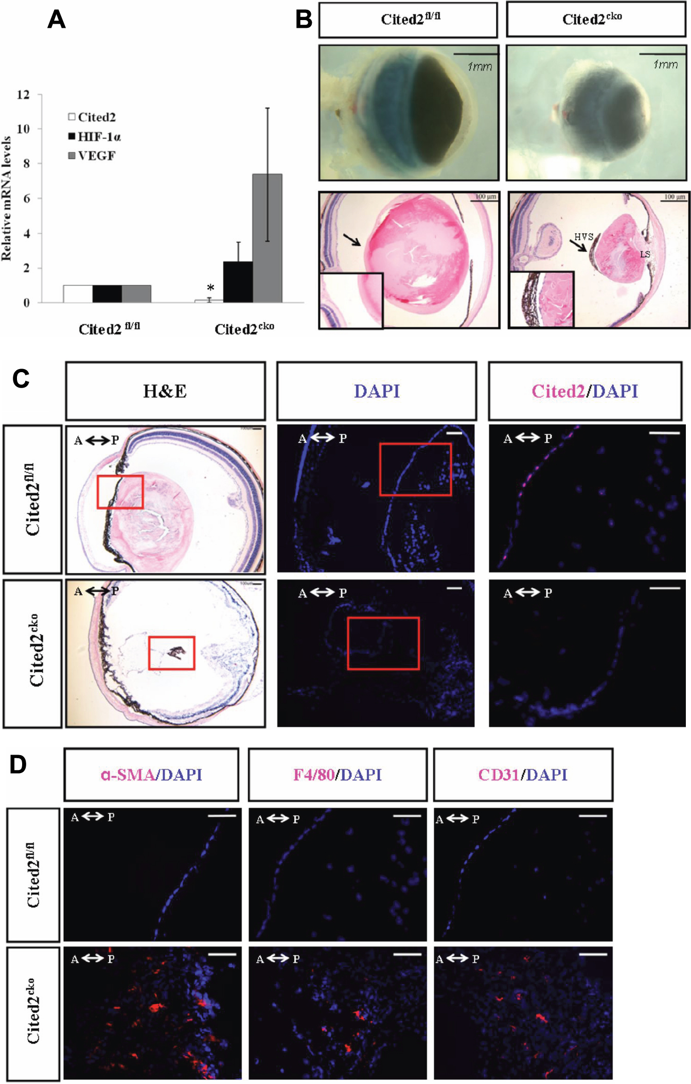
 Figure 1
of Huang, Mol Vis 2012; 18:1260-1270.
Figure 1
of Huang, Mol Vis 2012; 18:1260-1270.