Figure 8. Rod outer segment
phagocytosis by macrophage cells. A: Confocal
micrographs of macrophage cells incubated with FITC-modified
HNE-ROSs at 1 h (top, second) and at 24 h (top, third), and
bleached rod outer segments (ROSs) at 24 h (top, right).
FITC-ROSs co-localized well with macrophage cells that stained
with a fluorescent acidotropic probe of Lysotracker Red, which
show as yellow spots in microphotographs. There was more
co-localization in cells fed with HNE-ROSs than in cells fed
with bleached-ROSs at 24 h. B: Color channel separation
for the cells fed with HNE-modified ROSs at 24 h. Scale bar=10
μm.
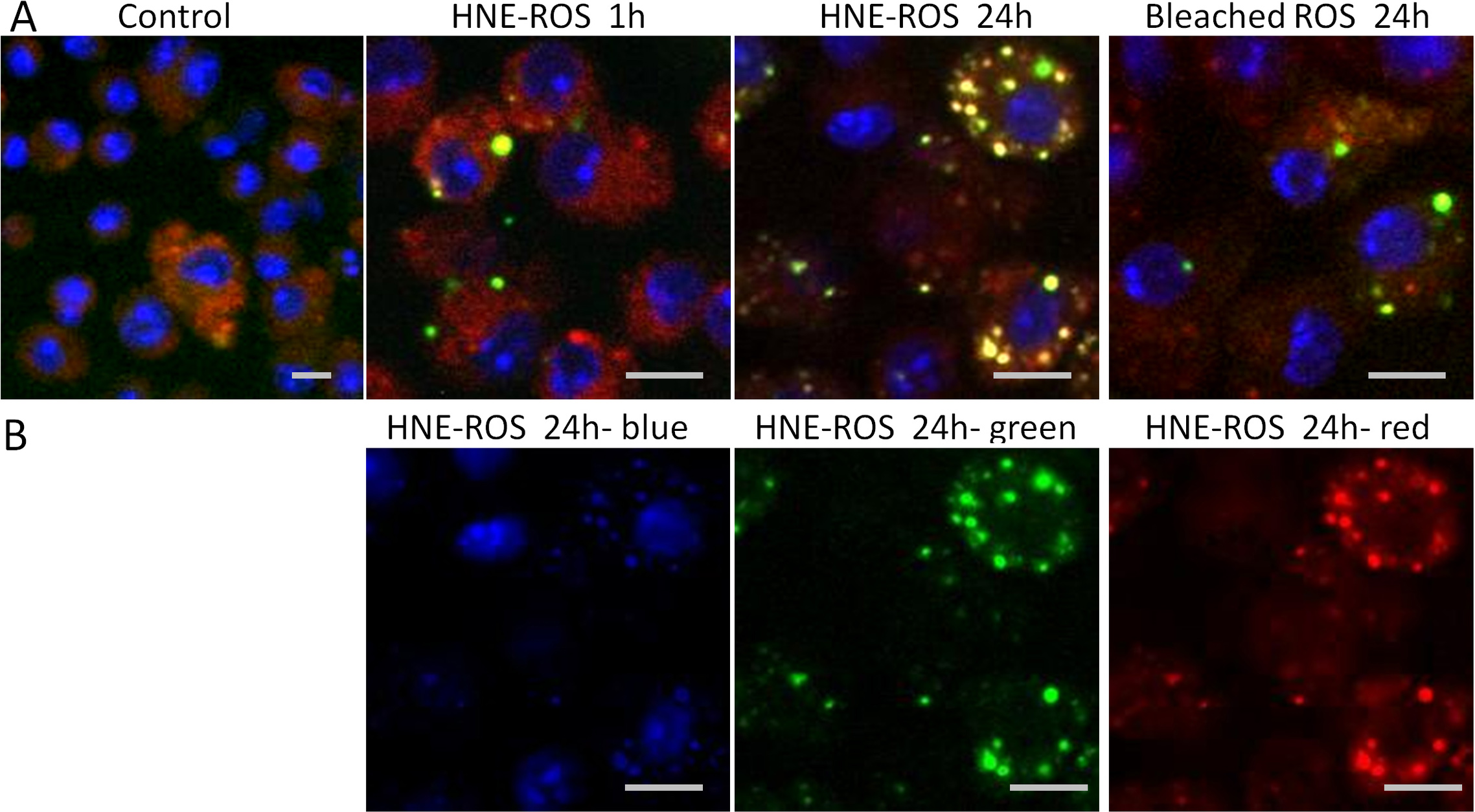
 Figure 8
of Lei, Mol Vis 2012; 18:103-113.
Figure 8
of Lei, Mol Vis 2012; 18:103-113.