Figure 4. Transmission electron
microscopy of microglial cells after 4 day feeding with ROSs. A:
Electron micrographs of microglial cells (control). B:
Electron micrograph of microglial cells after 4 day feeding with
bleached ROSs. Magnification: 6,000×. A higher magnification of
the intracellular inclusion body region is presented in the
micrograph below (magnification 26,000×). C. Electron
micrograph of microglial cells after 4 day feeding with
unbleached ROSs. Magnification: 4,200×. A higher magnification
of the intracellular inclusion body region is presented in the
micrograph below (magnification 43,000×). White arrows indicate
intracellular inclusion bodies in the higher magnification
photographs in B and C.
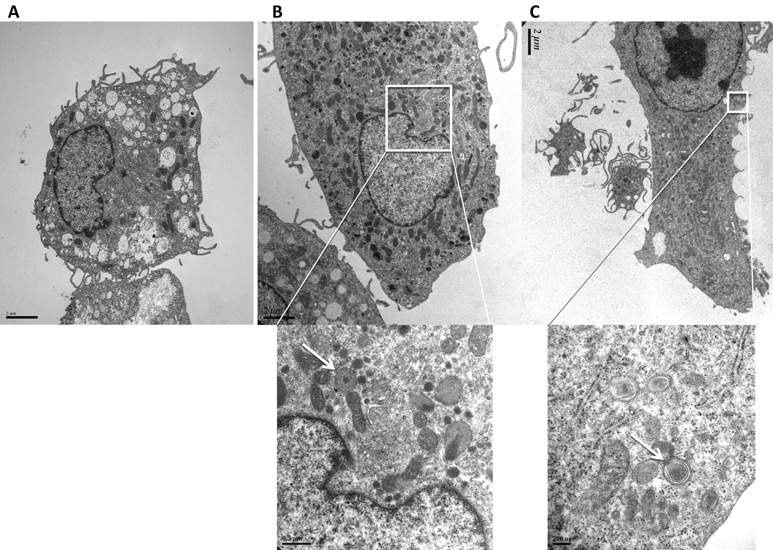
 Figure 4
of Lei, Mol Vis 2012; 18:103-113.
Figure 4
of Lei, Mol Vis 2012; 18:103-113.