Figure 3. Transfection with targeting
siRNAs suppressed TβRII protein expression by immunofluorescence
staining. Untreated SRA01/04 cells (A-C), SRA01/04
cells treated with 80 nM scrambled siRNA for 48 h (D-F),
or treated with 80 nM hT3 (G-I) were observed
under immunofluorescence microscope. Staining of nuclei with
Hoechst 33342 (blue) is shown in A, D, and G.
TβRII expression is shown in B, E, and H.
Compared with the control cells (B and E), TβRII
staining of cells treated with hT3 siRNA was weaker (H).
The morphology after the siRNA treatment were somewhat altered.
Note the exclusively cytoplasmatic localization of TβRII
expression (C, F, and I). Bar: 25 μm.
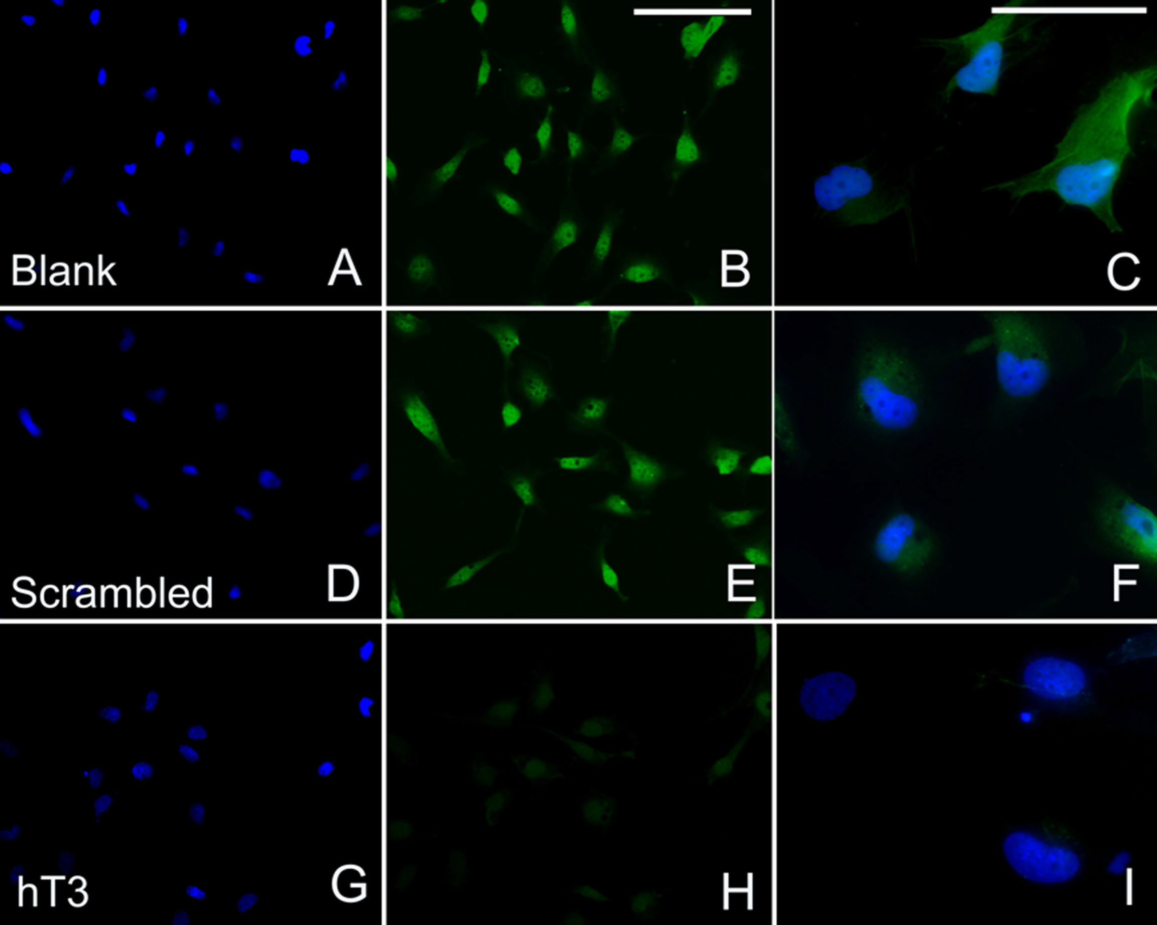
 Figure 3
of Zheng, Mol Vis 2012; 18:1238-1246.
Figure 3
of Zheng, Mol Vis 2012; 18:1238-1246.