Figure 2. Histology of corneas on
days 1, 3, 7, 14, and 30 post–Candida albicans keratitis
induction. Five corneas belonging to each Candida albicans
keratitis (CaK) group were serially sectioned at each time point
and adjacent sections were stained using the hematoxylin and
eosin (H&E) and periodic acid–Schiff (PAS) methods. H&E
staining was mainly used for examination of the cellular
distribution and gross structure of the cornea, and PAS staining
for revealing the distribution of fungi. One representative
section from each group at each time point is shown. A:
Structures of infected corneas changed significantly with time.
B: Lasting infiltration in corneas of both sham and
infected C57BL/6 mice, but only transient infiltration in Balb/c
corneas as indicated for day 30.
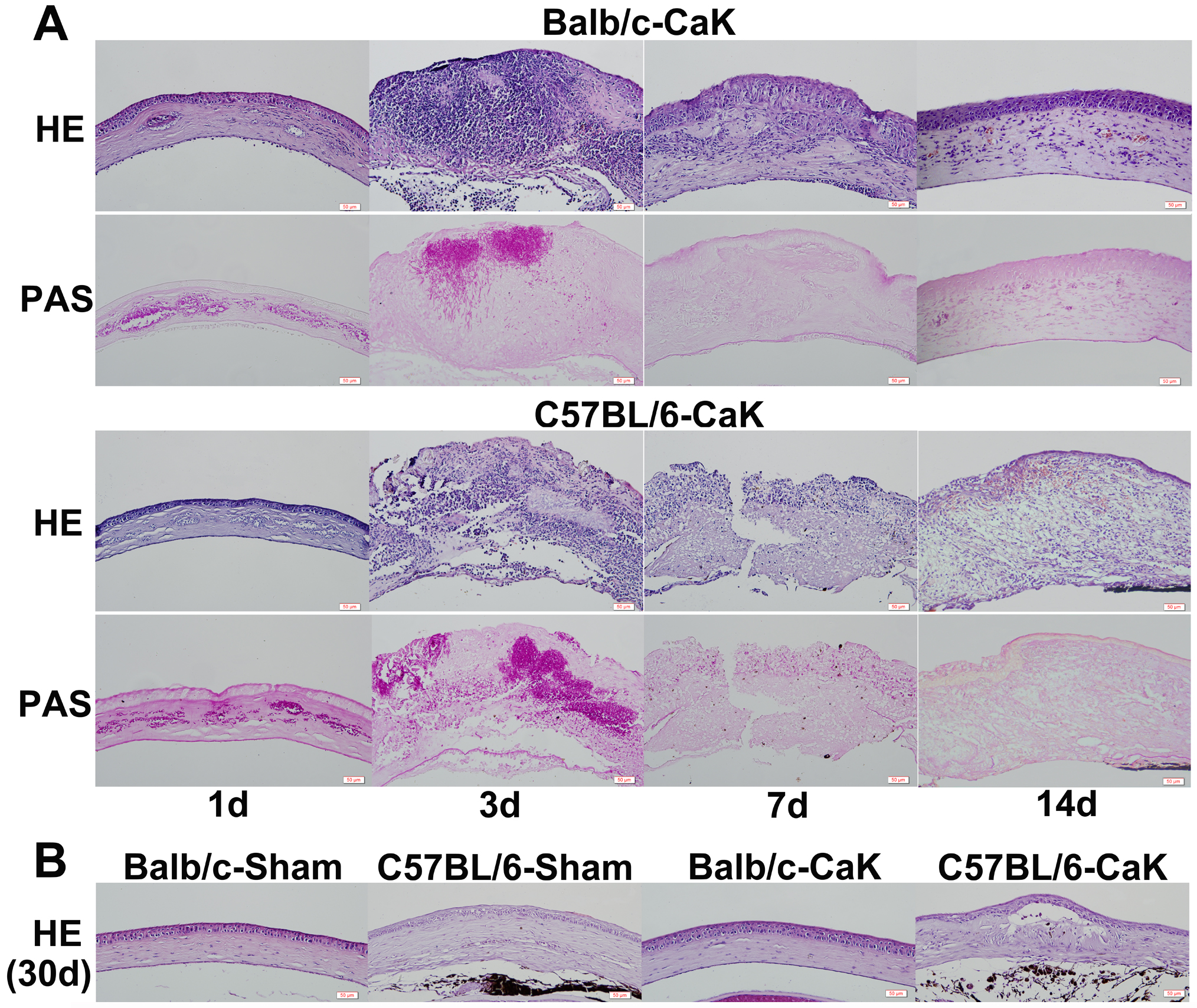
 Figure 2
of Zou, Mol Vis 2012; 18:1215-1225.
Figure 2
of Zou, Mol Vis 2012; 18:1215-1225.