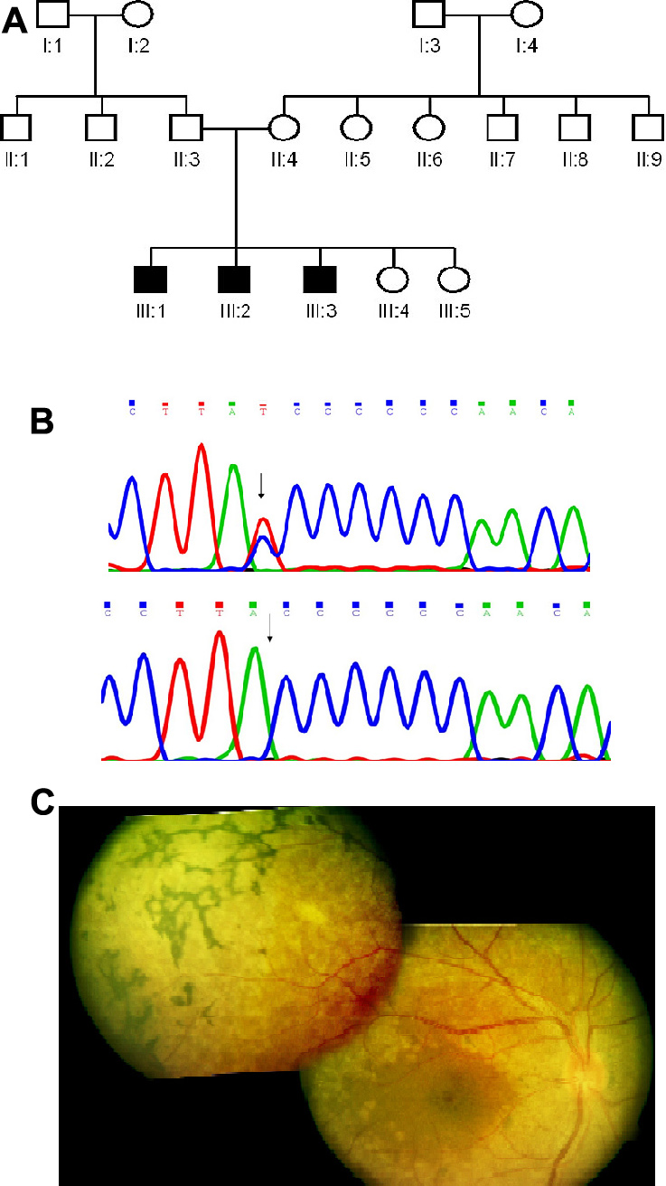
Figure 3. Molecular and clinical details of patient from Family C with a mutation in the
MFRP gene.
A: The family pedigree is shown.
B: Sequence of
MFRP gene in normal control (top panel) and in patient C-1 (bottom panel). The arrows in the top and bottom panels respectively,
mark the SNP c.492C>T (
rs36015759) and the position of the single base deletion in patient C-1.
C: Fundus montage of the right eye of patient C-1 (aged 21 years) from Family C with an
MFRP gene mutation showing perifoveal pigment deposits, relative parafoveal sparing, diffuse extensive graying of retina with
white flecks extending from arcades to the peripheral retina, and the presence of a peripheral reticular, bone corpuscular
type of pigmentary retinopathy. There is not much disc pallor or arterial narrowing.
 Figure 3 of
Kannabiran, Mol Vis 2012; 18:1165-1174.
Figure 3 of
Kannabiran, Mol Vis 2012; 18:1165-1174.