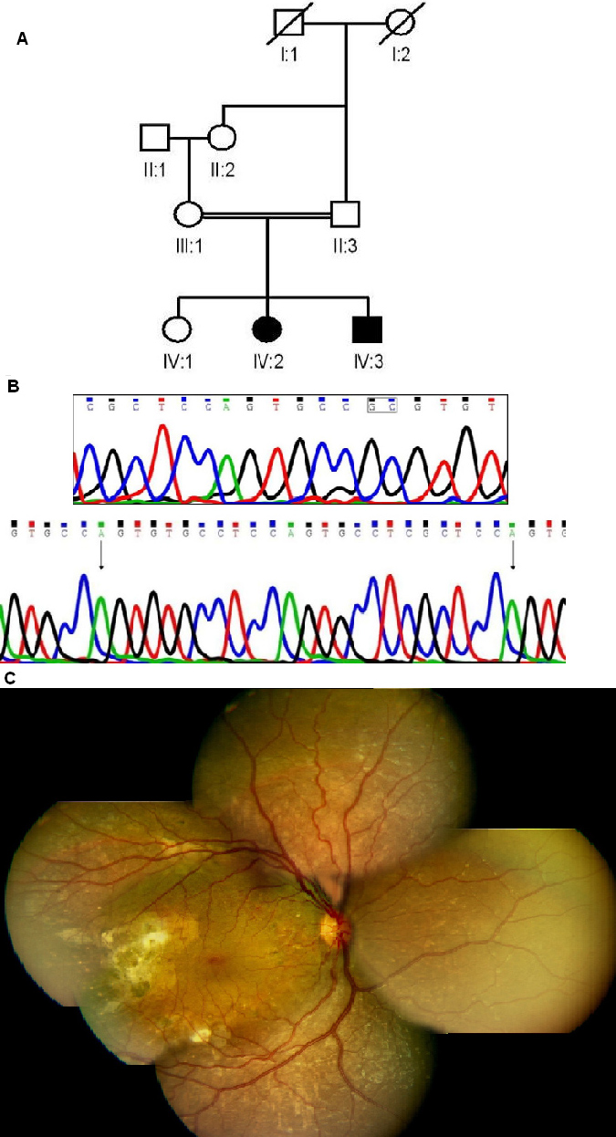
Figure 2. Molecular and clinical details of a patient from Family B with a mutation in the
NR2E3 gene.
A: Pedigree is shown (explanation of symbols as in
Figure 1).
B: Sequence of the
NR2E3 gene in normal control (top) and patient B2 with homozygous deletion+insertion (bottom). The dinucleotide undergoing deletion
is boxed in top panel. The arrows in the bottom panel mark the inserted sequence.
C: Fundus montage of right eye of patient B-2 (aged 10 years) with
NR2E3 mutation showing peripheral graying of retina with white flecks due to RPE atrophy with macular sparing with hardly any disc
or arterial changes. The right temporal retina had unexplained sub-retinal scarring/gliosis and no obvious bone corpuscular
pigment migration at this age.
 Figure 2 of
Kannabiran, Mol Vis 2012; 18:1165-1174.
Figure 2 of
Kannabiran, Mol Vis 2012; 18:1165-1174.