Figure 6. Western blot analysis of GRP78/BiP. HeLa cells were harvested at 12 (A), 24 (B), and 48 (C) h after transfection with the pcTGFBI-WT-myc (WT), pcTGFBI-T538P-myc (T538P), and pcTGFBI-R555W-myc (R555W) plasmids. The
proteins were separated on 8% SDS-polyacrylamide gels, transferred, and probed with an anti-GRP 78 antibody. The amount of
β-actin was analyzed with an anti-β-actin antibody to ensure that equal amounts of protein were loaded in each lane for electrophoresis.
The intensity ratio was calculated by the intensity of the GRP78 band divided by the intensity of the β-actin band (D). No significant differences were found between the mutant and wild-type groups at 12 and 48 h (p>0.05). The intensity ratio
of the T538P group was slightly lower than the wild-type group at 24 h (p=0.003). This transient fluctuation returned to the
same level as the wild-type group after 48 h. The expression of GRP78/BiP at 48 h was significantly higher than that at 12
and 24 h (p<0.05). In the positive control group, GRP78/BiP expression was significantly increased in the tunicamycin-treated
cells compared with the untreated cells (E).
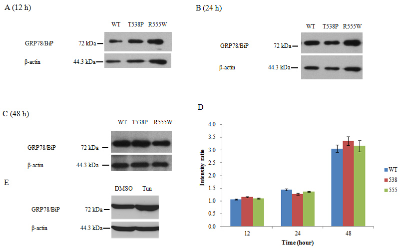
 Figure 6 of
Zhu, Mol Vis 2012; 18:1156-1164.
Figure 6 of
Zhu, Mol Vis 2012; 18:1156-1164.