Figure 4. Western blot analysis of the recombinant wild-type and mutant proteins. HCE and HeLa cells (A, B: HCE cell; C, D: HeLa cells) were transfected with wild-type (lane 2) and two mutant TGFBI plasmids (lanes 3 and 4) or were not transfected
plasmid (lane 1). The culture medium was replaced with serum-free medium 48 h post-transfection. Another 48 h later, the serum-free
media from overexpressed cells were collected and concentrated. A and C: 65 µg protein was loaded into each well. The TGFBI-T538P-myc and TGFBI-R555W-myc protein were detected with an anti-c-myc-tag
antibody (lanes 3 and 4), while the wild-type TGFBI-myc protein was not (lane 2). B and D: 65 µg protein was loaded on to each well and detected with an anti-TGFBIp antibody. The wild-type and mutant TGFBI-myc fusion
proteins were detected as two bands of different intensities (lanes 2–4). Endogenous TGFBIp is shown in the control lane (lane
1). The stronger bands in lanes 3 and 4 represent full-length mutant TGFBI-myc proteins, while the stronger band in lane 2
represents the wild-type TGFBI-myc protein that lacks its C-terminus. There was an approximate 10 kDa difference between the
full-length and cleaved proteins.
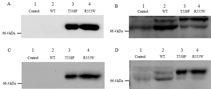
 Figure 4 of
Zhu, Mol Vis 2012; 18:1156-1164.
Figure 4 of
Zhu, Mol Vis 2012; 18:1156-1164.