Figure 2. Schematic representation of the recombinant mutant TGFBI proteins and the principal of the two-sequential PCR site-directed
mutagenesis technique. A: S: signal sequence, EMI: cysteine-rich EMI domain, I to IV: four FAS1 domains, RGD: Arg-Gly-Asp domain, Myc: myc tag protein,
TGFBI-T538P-I: bp 1 to 1613 (the 538 mutant site), TGFBI-R555W-I: bp 1 to 1663 (the 555 mutant site), TGFBI-T538P/R555W-II: from the mutant site to the 3′ end of the sequence. B: The rectangle represents the template DNA, the circle in the rectangle represents the mutant site, and the arrow represents
the primer. In the first PCR, TGFBI-T538P/ R555W-I were amplified with the TGFBI-WT-F and TGFBI-T538P/ R555W-R primers. TGFBI-T538P/ R555W-II were amplified with the TGFBI-T538P/ R555W-F and TGFBI-WT-R primers. In the second PCR, full-length TGFBI-T538P/ R555W were amplified with the TGFBI-WT-F and TGFBI-WT-R primers.
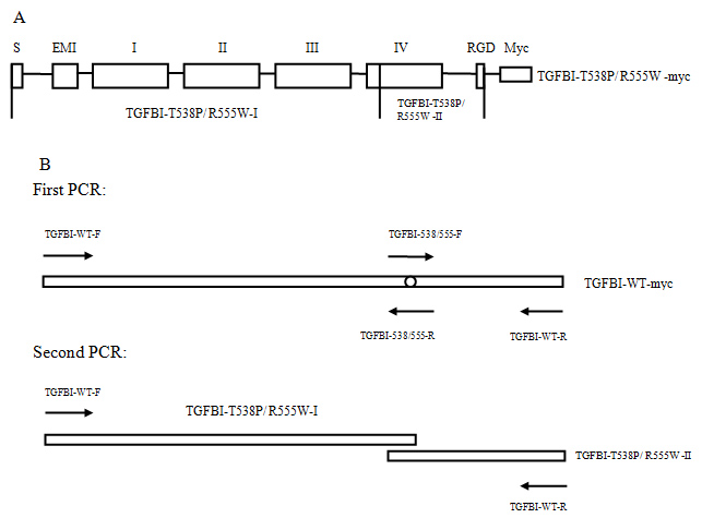
 Figure 2 of
Zhu, Mol Vis 2012; 18:1156-1164.
Figure 2 of
Zhu, Mol Vis 2012; 18:1156-1164.