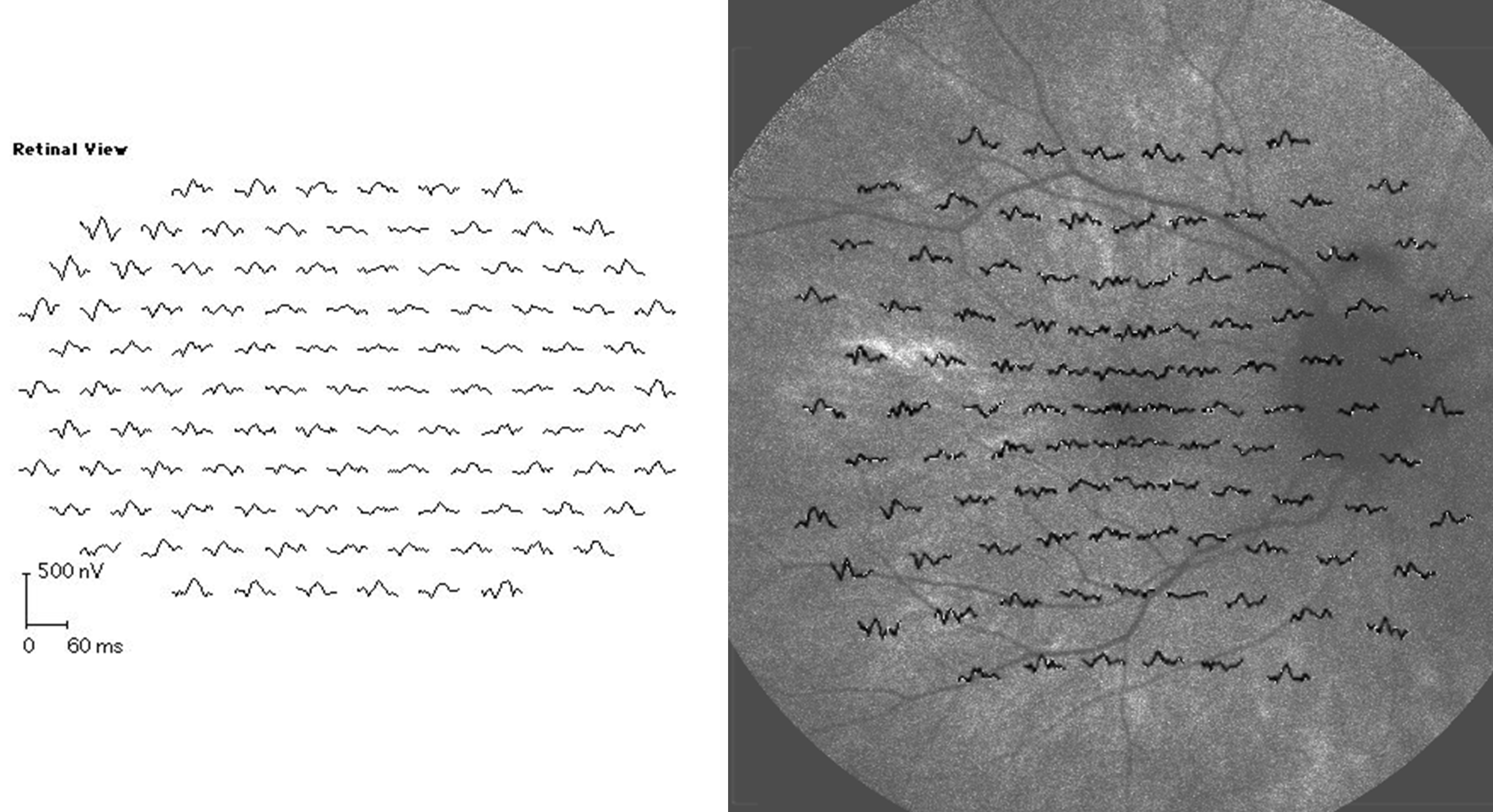
Figure 7. Multifocal
electroretinography (left panel) and overlay of multifocal
electroretinography focal responses on fundus autofluorescence
photography (right panel) in the 33-year-old female carrier of
X-linked retinitis pigmentosa whose fundus is presented in
Figure 6.
Patchy retinal dysfunction compatible with random X
inactivation, and a patchy variability in fundus
autofluorescence intensity is demonstrated. Areas of remaining
autofluorescence seem to correspond to areas of remaining
responses by multifocal electroretinography. The following
abbreviations apply: ms is short for millisecond and nV is short
for nanovolt.
 Figure 7
of Schatz, Mol Vis 2012; 18:1147-1155.
Figure 7
of Schatz, Mol Vis 2012; 18:1147-1155.