Figure 7. Expression and localization
of CTRP5 in the pig eye. A: Expression of pCTRP5
was studied by qRT–PCR using total mRNA extracted from different
tissues of a 222-day-old pig eye. The pCTRP5 expression
is presented as bars using an arbitrary scale on the y
axis. Values are presented as mean (±SEM) of three independent
observations after normalization with the control gene (HGPRT) B:
western blot analysis of CTRP5 protein extracted from a 222 days
pig. Retina (lane 1), optic nerve (lane 2), lens (lane 3), RPE
(lane 4), ciliary body (lane 5), Choroid (lane 6). Detection
with an anti-CTRP5 antibody shows significant expression of
pCTRP5 protein in the RPE with an expected size molecular weight
of approximately 31 kDa. C: Localization of CTRP5
in retinal sections as evaluated by IHC analysis of retinal
sections with human monoclonal anti-CTRP5 antibody and Alexa
Fluor 488 staining (green, arrows), nuclei (*) stained with DAPI
(blue). D: Localization of CTRP5 in the ciliary body as
shown by IHC with human monoclonal anti-CTRP5 antibody and Alexa
Fluor 555 (red, arrows), nuclei stained with DAPI (blue).
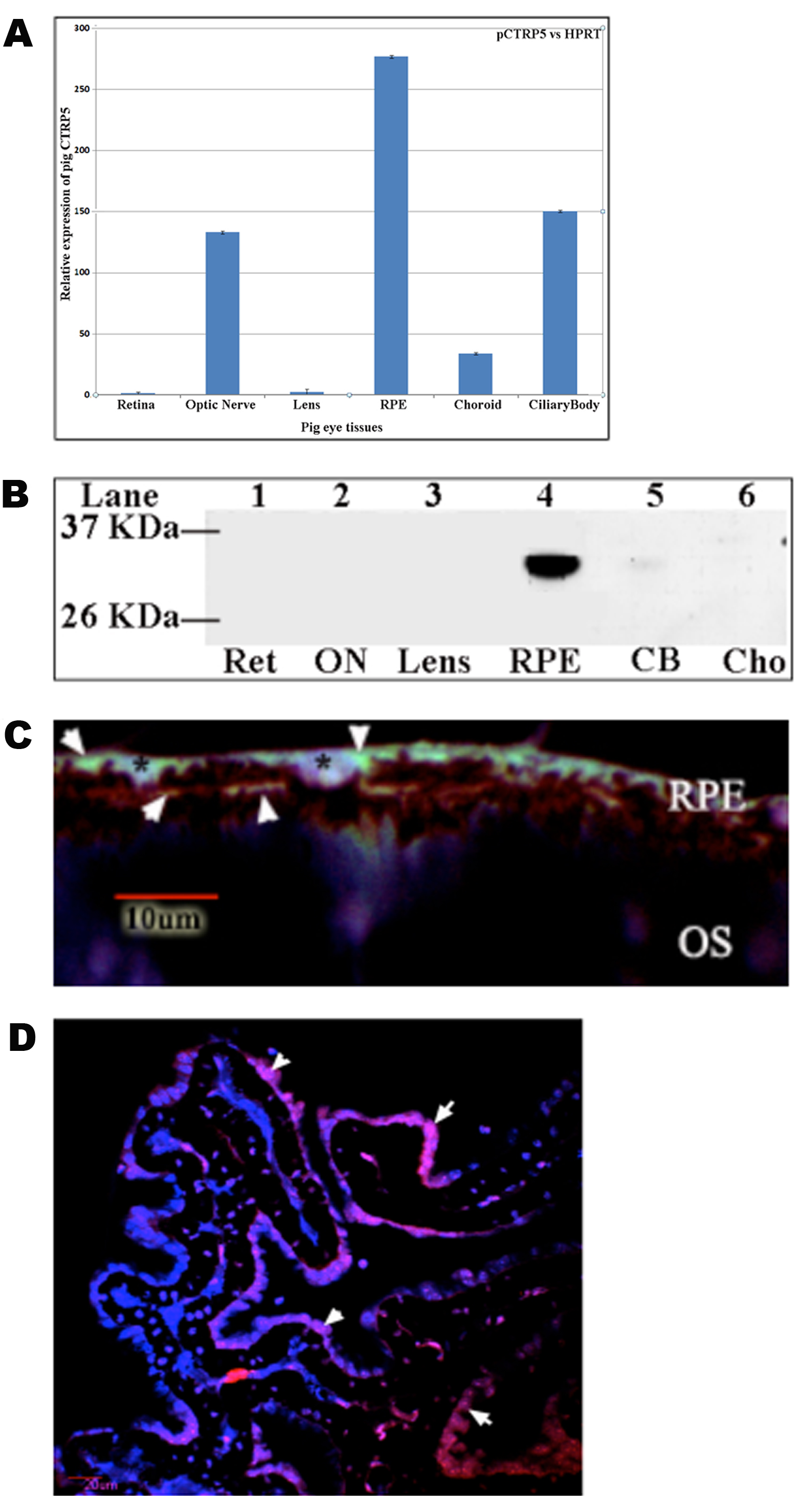
 Figure 7
of Sommer, Mol Vis 2012; 18:92-102.
Figure 7
of Sommer, Mol Vis 2012; 18:92-102.