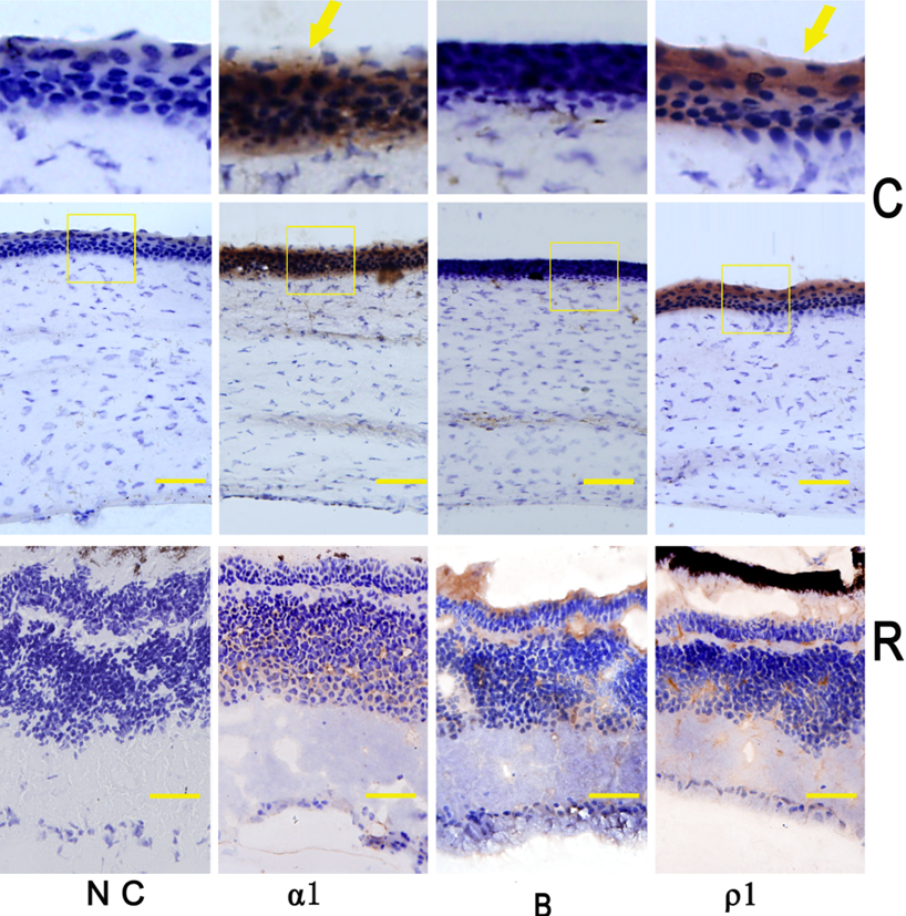
Figure 3. Alpha
1 GABA
A
(α1), GABA
B (B) and rho
1 GABA
C
(ρ1) receptor protein distribution in chick cornea (C) and
retina (R). With antibodies for alpha
1 GABA
A,
GABA
B and rho
1 GABA
C receptors,
immunoreactivity was observed in corneal epithelium cell for
alpha
1 GABA
A (α1) and rho
1 GABA
C
(ρ1; yellow arrow), but not for GABA
B receptor (B) in
the cornea (C). In the retina (R), immunoreactivity was found in
the inner plexiform layer, outer plexiform layer, inner nuclear
layer, and ganglion cell layer for alpha
1 GABA
A
(α1), GABA
B (B) and rho
1 GABA
C
(ρ1) receptors, corresponding to previous reports [
6-
11,
15].
There was essentially no immunoreactivity observed for the
negative controls (N.C). The scale bar is 10 µm. Photographs
were taken at 40× magnification.
 Figure 3
of Cheng, Mol Vis 2012; 18:1107-1114.
Figure 3
of Cheng, Mol Vis 2012; 18:1107-1114.