Figure 7. Retinal architecture of
Aca23 and WT. Representative cross-section images of the retinas
of Aca23 and WT mice before treatment. The Aca23 retina (A)
has no differences in retinal layer thicknesses from WT (B;
stained with Toluidine Blue; scale bar=20 µm).
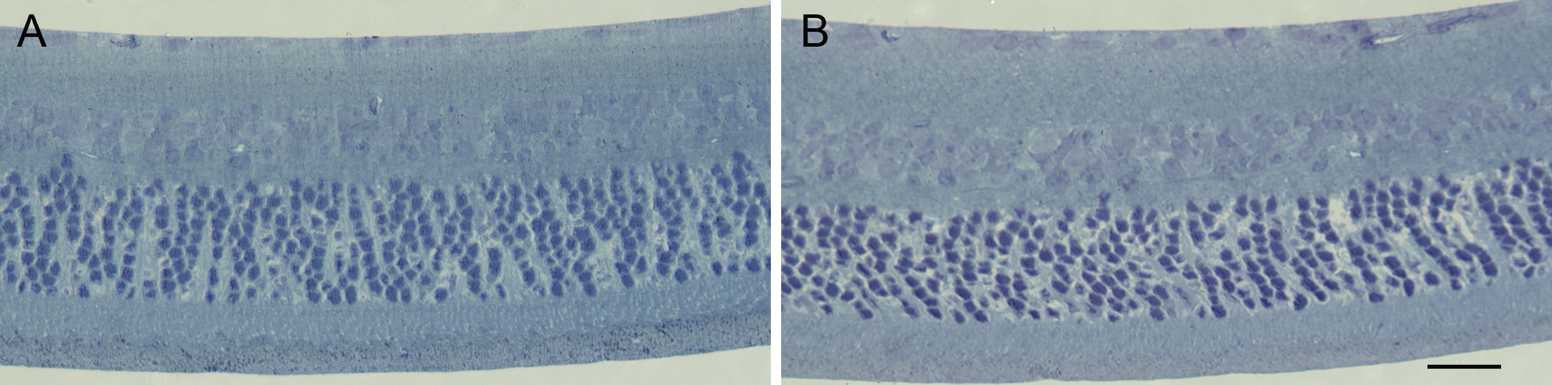
 Figure 7
of Steinhart, Mol Vis 2012; 18:1093-1106.
Figure 7
of Steinhart, Mol Vis 2012; 18:1093-1106.