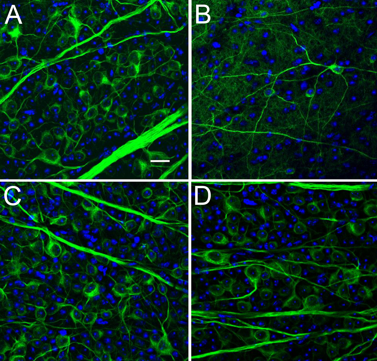
Figure 6. RGC loss in retinal whole mount. Representative images of the RGC layer of retinal wholemounts stained with DAPI (blue) and
anti-β-tubulin (green). A: An untreated WT control retina with nuclei of all cells including RGC, amacrines and others labeled blue with DAPI. The
cytoplasm, dendrites and axons of RGC labeled green for tubulin. B: Glaucoma retina from WT animal with substantial loss of RGC. C: An untreated Aca23 control retina is similar to untreated WT. D: Aca23 retina after glaucoma, showing one of the retinas with modest loss of RGC compared to untreated Aca23 (C) or WT (B), but less than that of treated WT (B). (scale bar=20 µm).

 Figure 6 of
Steinhart, Mol Vis 2012; 18:1093-1106.
  Figure 6 of
Steinhart, Mol Vis 2012; 18:1093-1106.