Figure 5. Axon loss in optic nerve cross section. Representative images of optic nerve cross sections stained with Toluidine Blue. A: The untreated nerve from a WT animal shows no damage. B: Six weeks post intracameral microbead injection, the axons of RGCs show significant loss of axons. C: An untreated Aca23 nerve resembles that of an untreated WT animal. However after microbead injection (D), the treated Aca23 nerve does not show similar damage to the WT treated nerve (C). (scale bar=30 µm).
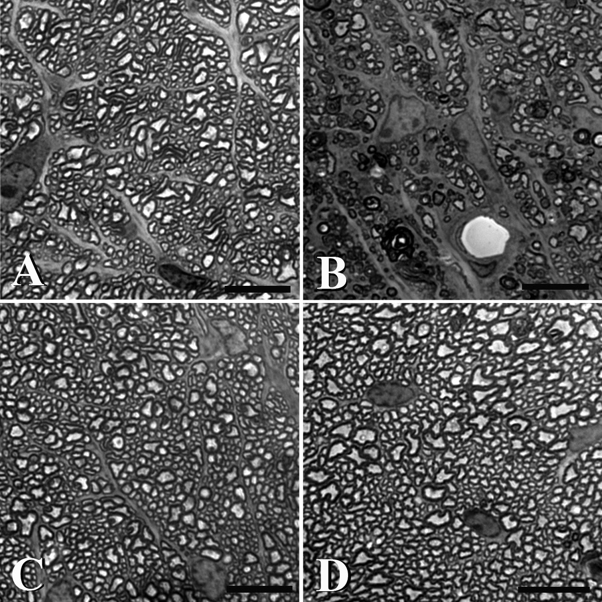
 Figure 5 of
Steinhart, Mol Vis 2012; 18:1093-1106.
Figure 5 of
Steinhart, Mol Vis 2012; 18:1093-1106.