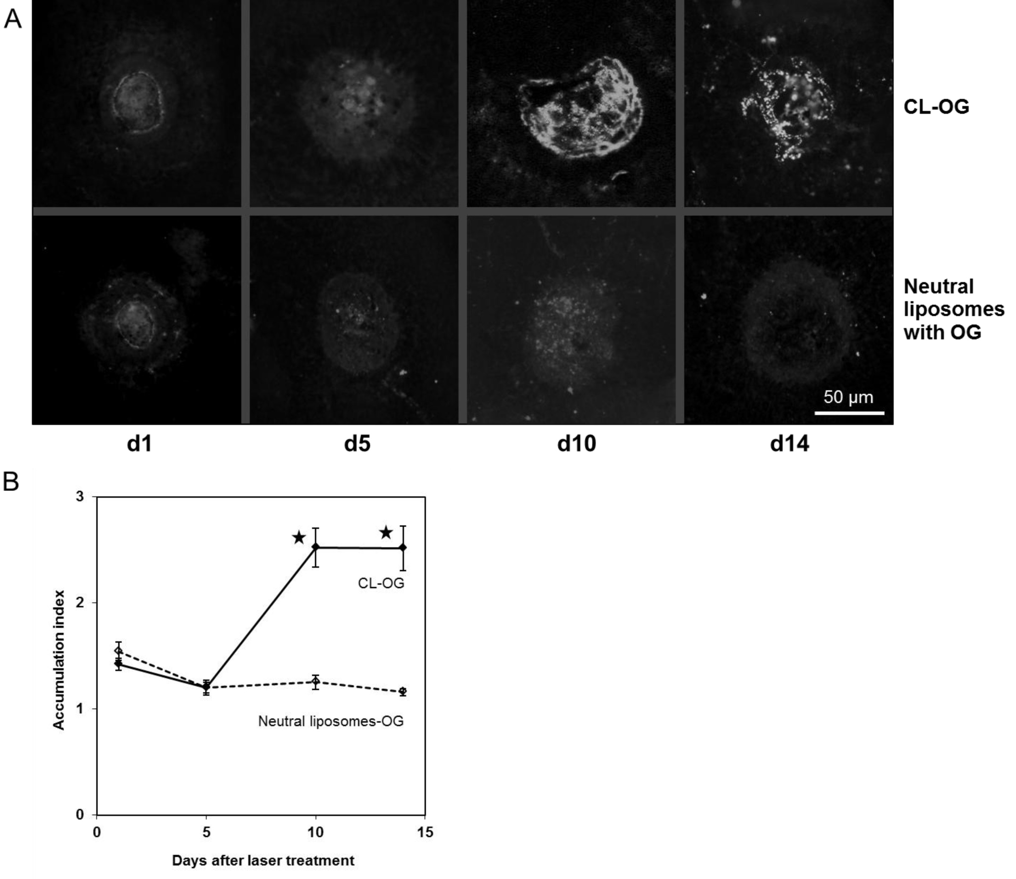
Figure 4. Accumulation of cationic
and neutral liposomes was observed in flatmount preparations.
After the scanning laser ophthalmoscope (SLO) images described
in
Figure 3
were taken, choroidal-scleral flatmounts were prepared from the
mice treated with cationic liposomes (CL)-Oregon green (OG) or
neutral liposomes labeled with OG. Images were taken using
fluorescence microscopy. Representative ones are shown in
A.
They were evaluated the same way as the SLO images (
B).
Values for CL-OG at d10 and d14 were significantly higher
compared to those of neutral liposomes. The results confirmed
that the kinetics were the same as for the SLO images. Data are
means obtained from five mice. Error bars indicate SEM, and
asterisks indicate statistical significance (p<0.05 as
compared to d1).
 Figure 4
of Hua, Mol Vis 2012; 18:1045-1054.
Figure 4
of Hua, Mol Vis 2012; 18:1045-1054.