Figure 3. Schematic diagram showing a
possible consequence from the c.163–1G→T transversion in FRMD7.
A: Schematic diagram of wild type FRMD7. B:
The mutant destroys the original splicing acceptor on the 3′
side of intron 2, and thus causes exon 3 (bases in green font)
skipping and creates a truncated protein of 64 amino acids. C:
Another altered splicing introduces a new stop codon and creates
a truncated protein of 59 amino acids.
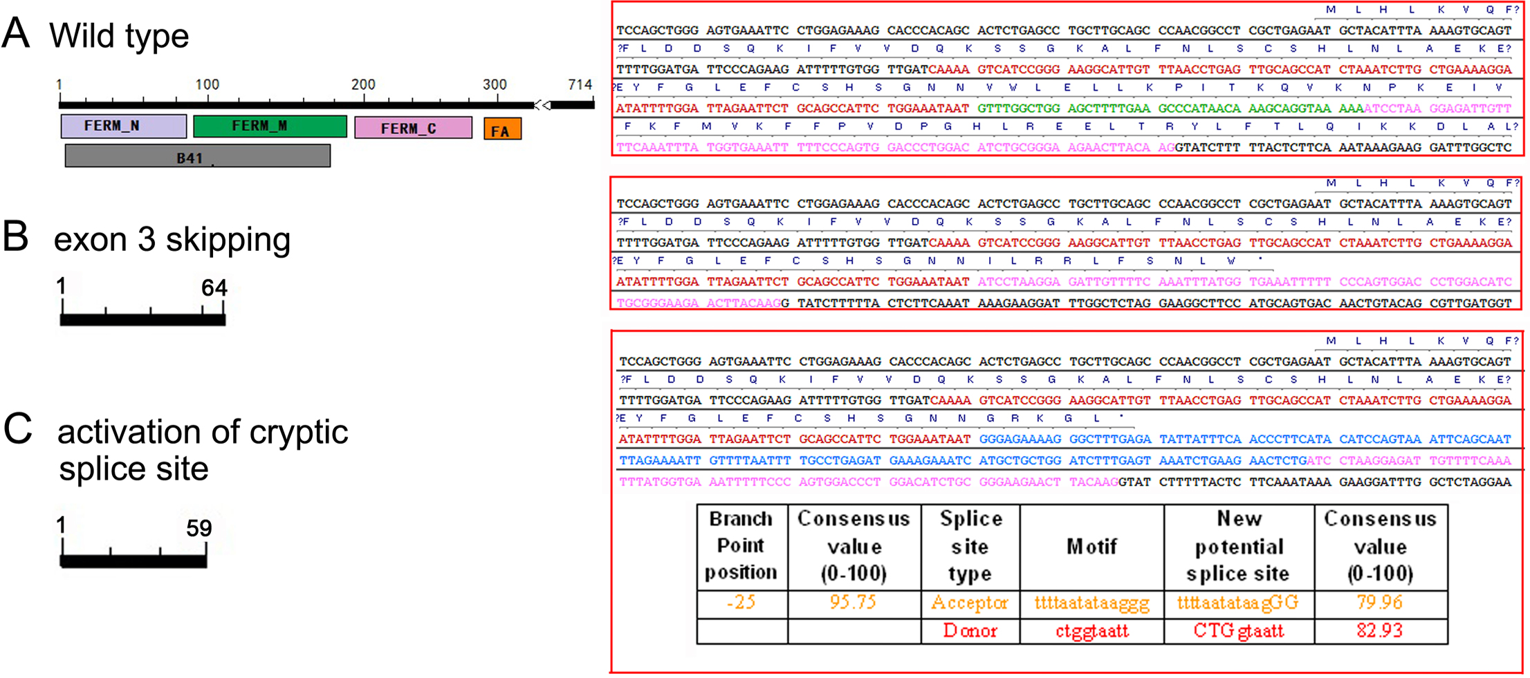
 Figure 3
of Hu, Mol Vis 2012; 18:87-91.
Figure 3
of Hu, Mol Vis 2012; 18:87-91.