Figure 2. Full-field
electroretinograms (ERGs) recorded according to the
International Society for Clinical Electrophysiology of Vision
(ISCEV) standards protocol in this case. The rod, combined
rod-cone, cone, oscillatory potentials, and 30-Hz flicker
full-field ERGs are shown. The results of full-field ERGs are
within the normal limits in this case.
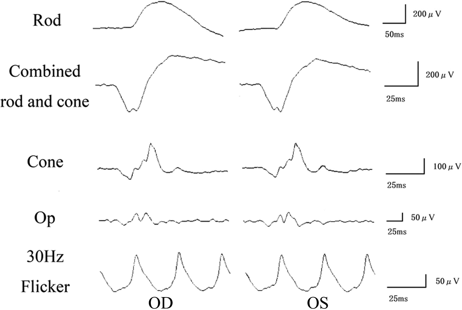
 Figure 2
of Kabuto, Mol Vis 2012; 18:1031-1039.
Figure 2
of Kabuto, Mol Vis 2012; 18:1031-1039.