Figure 1 of
Kabuto, Mol Vis 2012; 18:1031-1039.
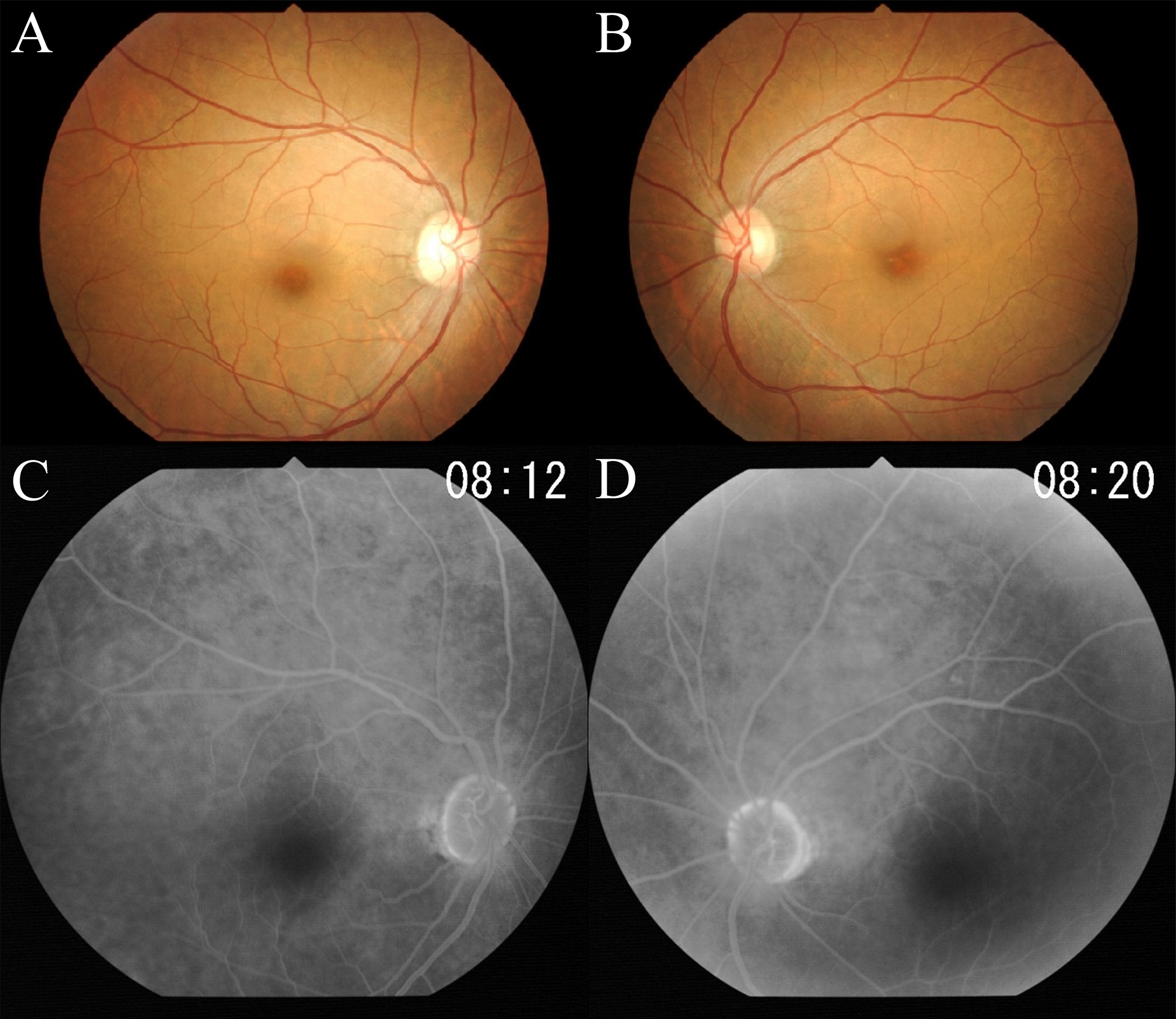
Figure 1.
Fundus photographs (
A
,
B
) and fluorescein angiograms (
C
,
D
) of this case showing no abnormal findings.
 Figure 1
of Kabuto, Mol Vis 2012; 18:1031-1039. Figure 1
of Kabuto, Mol Vis 2012; 18:1031-1039.
Figure 1
of Kabuto, Mol Vis 2012; 18:1031-1039. Figure 1
of Kabuto, Mol Vis 2012; 18:1031-1039.