Figure 3. Localization, retinal
histology, and thickness measurements for the Rdh13
knockout mice. A: Immunofluorescence localization of Rdh13
(green) was shown in 3-month-old Rdh13+/+ and
Rdh13−/− retina paraffin sections. B:
western blot analysis of RDH13 protein in wild-type (WT) and
homozygous mice revealed that there was no expression of Rdh13
in the retinas of Rdh13 knockout mice. C:
Semi-thin sections of WT and homozygous mice retinas revealed no
major differences in retinal histology at 3 and 10 months of
age. D: Transmission electron microscopy (TEM) of the
photoreceptor outer and inner segments and the outer nuclear
layer in Rdh13+/+ and Rdh13−/−
mice at 10 months of age revealed no apparent abnormalities. E:
The outer-plus-inner-segment and outer nuclear layer thickness
for Rdh13−/− and WT mice at the ages of 3
months and 10 months was valued. Values were mean±SD (n=5, each
group). There were no statistically significant differences
between the two genotypes at any distance point. ONH, optic
nerve head; RPE, retinal pigment epithelia; OS, outer segments;
IS, inner segments; ONL, outer nuclear layer; OPL, outer
plexiform layer; INL, inner nuclear layer; IPL, inner plexiform
layer; GCL, ganglion cell layer.
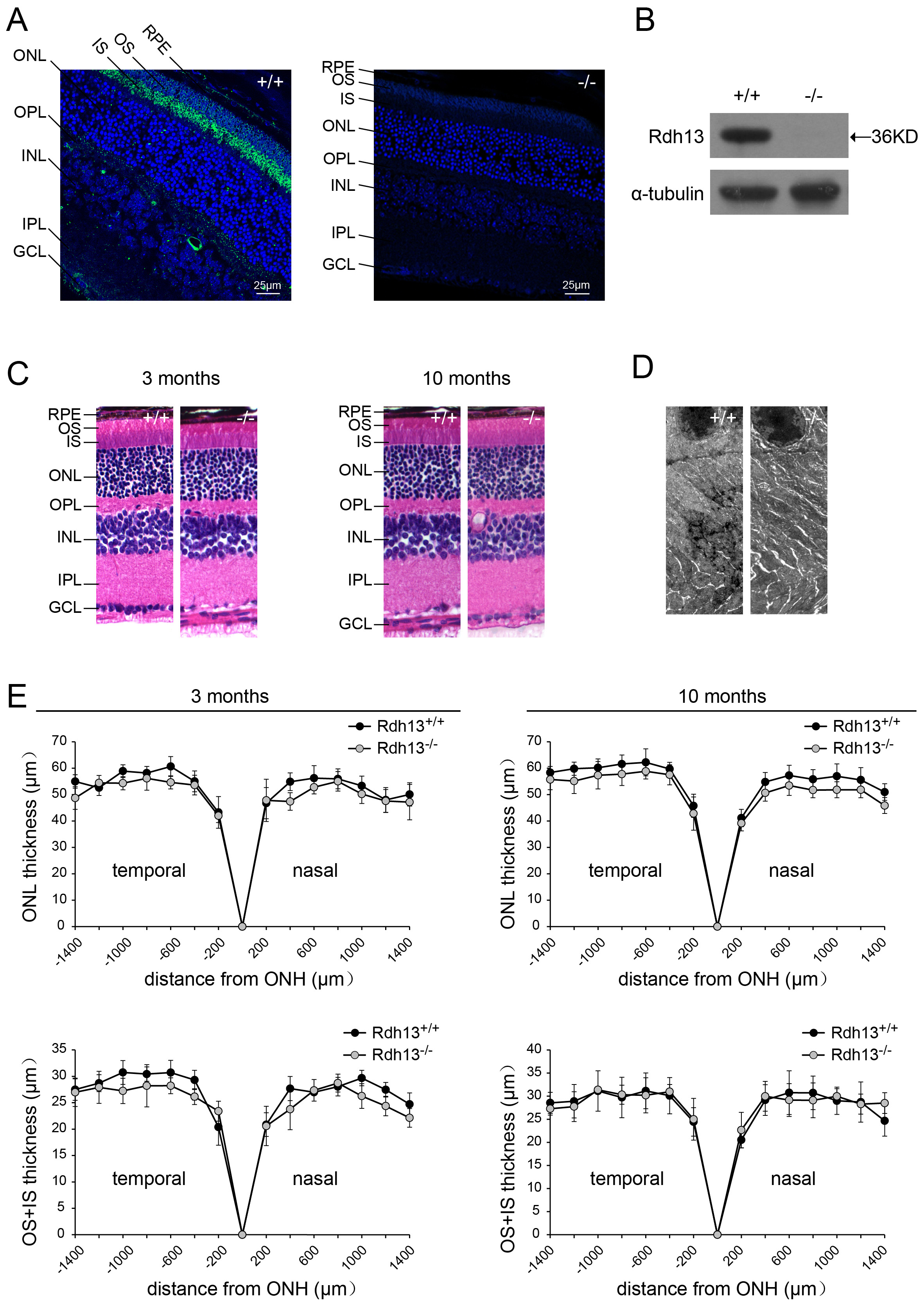
 Figure 3
of Wang, Mol Vis 2012; 18:1021-1030.
Figure 3
of Wang, Mol Vis 2012; 18:1021-1030.