Figure 2. Generation of Rdh13
knockout mice. A: This is the graphic representation of
the Rdh13 gene knockout strategy for the deletion of Rdh13
exons 2 and 3 in embryonic stem cells. Exons are shown in boxes.
The targeting vector was designed to delete exon 2 and exon 3.
The targeting vector contained a 2.9 kb 5′ arm and 3.2 kb 3′
arm. PGK-Neo and HSV-TK cassettes were used for positive and
negative selections, respectively. The genomic positive of the
PCR primers for genotyping are indicated by arrows. B:
Genomic DNA from ES cell clones was isolated and analyzed by
PCR. The successfully targeted embryonic stem cell DNA was
amplified into 6.2 kb and 3.5 kb products for the 5′ arm and 3′
arm, respectively. C: The genotype of Rdh13+/+,
Rdh13+/−, and Rdh13−/− mice
was detected by PCR. D: Rdh13 transcripts in
mouse liver from Rdh13+/+, Rdh13+/−,
and Rdh13−/− mice was analyzed by
reverse-transcription PCR. E: The expression pattern of
RDH13 protein in Rdh13+/+, Rdh13+/−,
and Rdh13−/− mouse liver was revealed by
western blot.
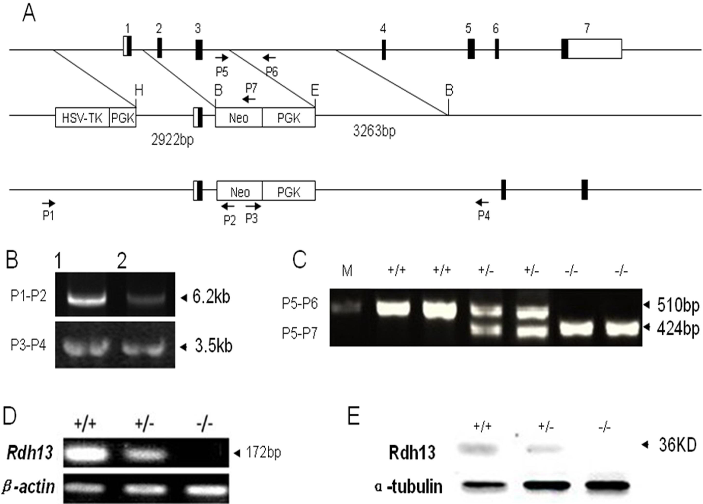
 Figure 2
of Wang, Mol Vis 2012; 18:1021-1030.
Figure 2
of Wang, Mol Vis 2012; 18:1021-1030.