Figure 6. Pirfenidone inhibited transforming growth factor-β1 (TGF)-β1-induced signal transduction. A: Human retinal pigment epithelial cells (ARPE-19) cells were incubated in the absence or presence of pirfenidone (500 mg/l)
for 1 h, then treated with TGF-β1 (10 μg/l) for varying time periods, and total cell lysates were subjected to immunoblot
analysis for phosphor-Smad and Smad2/3. The data shown are representative of three independent experiments. B: Nuclear extracts from the cells incubated in the absence or presence of pirfenidone (500 mg/l) for 1 h and then treated
with TGF-β1 (10 μg/l) for an additional 30 min were subjected to immunoblot analysis for Smad2/3. Poly (ADP-ribose) polymerase
(PARP) was used for a positive control for nuclear compartment; while α-tubulin was used for a positive control for cytosolic
fraction. C: Cells were incubated in the absence or presence of pirfenidone (500 mg/l) for 1 h, then treated with TGF-β1 (10 μg/l) for
30 min, and stained with antibody against phospho-specific Smad2/3 and secondary antibody conjugated with fluorescein isothiocyanate
(FITC; green). Nucleus was counter-stained with propidium iodide (red). Scale bar=20 μm. The data shown are representative
of three independent experiments.
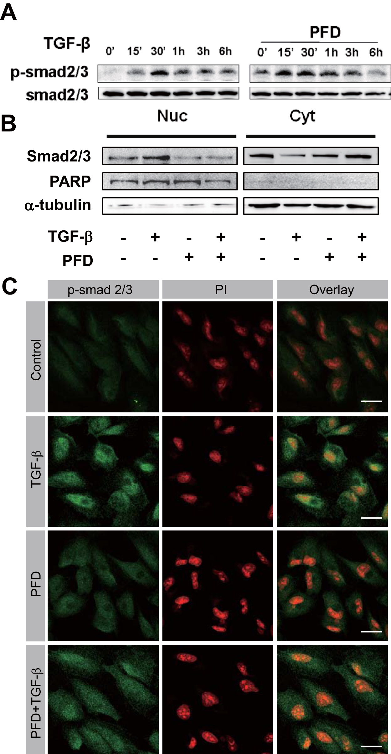
 Figure 6 of
Choi, Mol Vis 2012; 18:1010-1020.
Figure 6 of
Choi, Mol Vis 2012; 18:1010-1020.