Figure 1. Pirfenidone inhibited
transforming growth factor- β1 (TGF- β1)-induced morphological
changes and actin rearrangement in a human retinal pigment
epithelial cells, ARPE-19. A: ARPE-19 cells were
incubated in the absence or presence of pirfenidone (500 mg/l)
or hydroxyfasudil (10 μmol/l) for 1 h, treated with TGF-β1 (10
μg/l) for an additional 48 h, and visualized with phase contrast
microscopy. The data shown are representative of at least four
independent experiments. Magnification, 100×. Scale bar=20 μm. B:
Cells were incubated in the absence or presence of pirfenidone
(500 mg/l) or hydroxyfasudil (10 μmol/l) for 1 h, treated with
TGF-β1 (10 μg/l) for an additional 48 h, and stained with
rhodamine-labeled phalloidin for F-actin and fluorescein
isothiocyanate (FITC)-conjugated antibodies for N-cadherin. The
data shown are representative of at least three independent
experiments. Magnification, 400×. Scale bar=20 μm. C:
Cells were incubated in the absence or presence of pirfenidone
(500 mg/l) for 1 h and then treated with TGF-β1 (10 μg/l) for
varying time periods. The total cell lysates were subjected to
immunoblot analysis for phospho-cofilin, cofilin, and β-actin.
The data shown are representative of at least two independent
experiments. Control: untreated, PFD: pirfenidone, fasudil:
hydroxyfasudil.
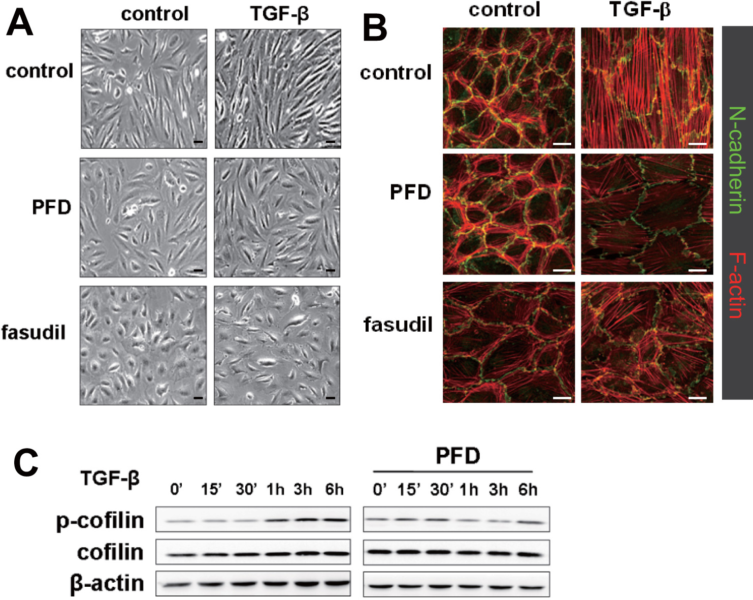
 Figure 1
of Choi, Mol Vis 2012; 18:1010-1020.
Figure 1
of Choi, Mol Vis 2012; 18:1010-1020.