Figure 2 of
Chen, Mol Vis 2012; 18:989-995.
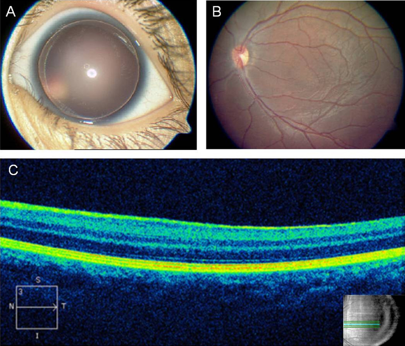
Figure 2.
Photos and images showing the clinical features of affected patients.
A
: Complete absence of iris in III-2 (OS).
B
: Foveal hypoplasia observed in III-2 (OS).
C
: A flat fovea in III-2 (OS) demonstrated by optical coherence tomography.
 Figure 2 of
Chen, Mol Vis 2012; 18:989-995.
Figure 2 of
Chen, Mol Vis 2012; 18:989-995.