Figure 4. Immunolocalization of AQP5
in mouse cornea. A: Schematic diagram of a mammalian
cornea showing the five layers. B: AQP5 localization
(green) in central corneal epithelial cells and stromal
keratocytes. C: AQP5 knockout mouse central corneal
epithelial cells and stromal keratocytes showing lack of
immunoreactivity. D: AQP5 localization (red) in the
limbal area of the cornea; the window in D is enlarged
and shown as E. E: AQP5 (red) in limbal stromal
keratocytes. F: AQP5 knockout mouse corneal stromal
keratocytes in the limbal area with no immunoreactivity. B,
C: FITC conjugated secondary antibody. D, E,
F: Texas Red conjugated secondary antibody; blue, nuclear
stain DAPI. Epi: epithelium; Str: stroma; En: endothelium;
arrows- antibody binding.
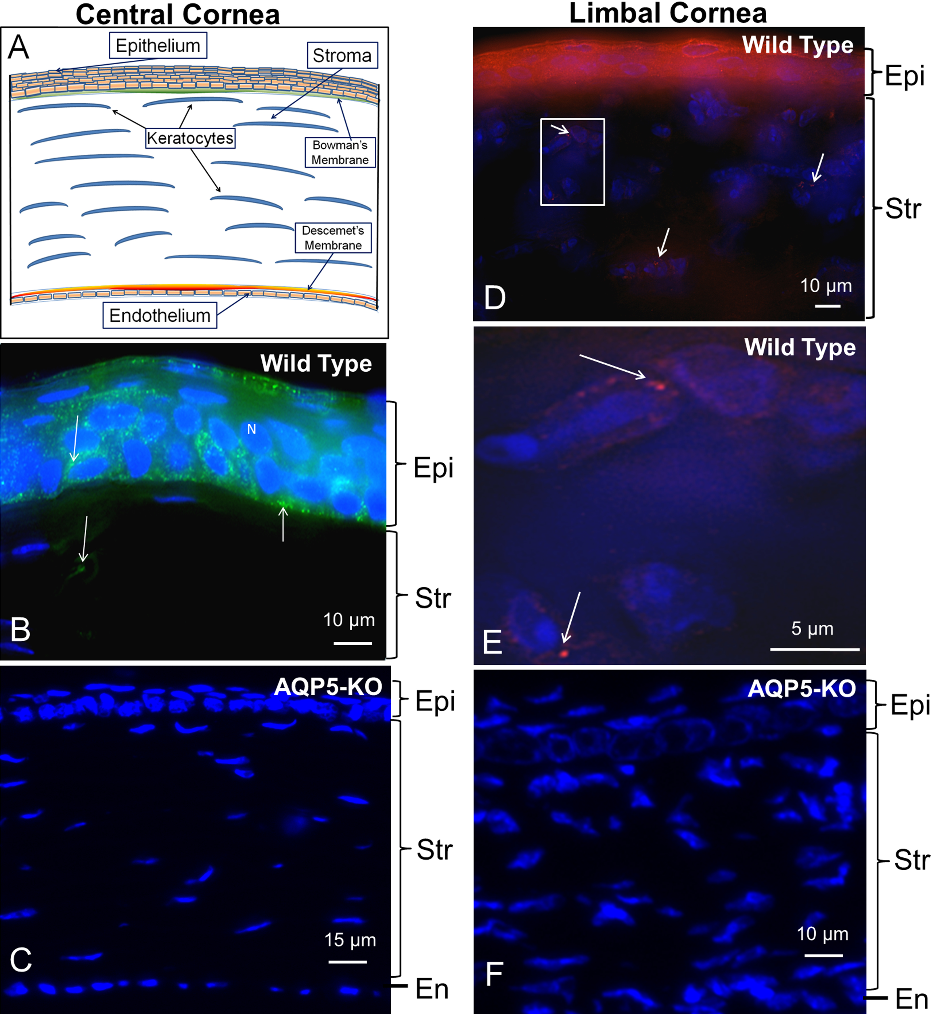
 Figure 4
of Kumari, Mol Vis 2012; 18:957-967.
Figure 4
of Kumari, Mol Vis 2012; 18:957-967.