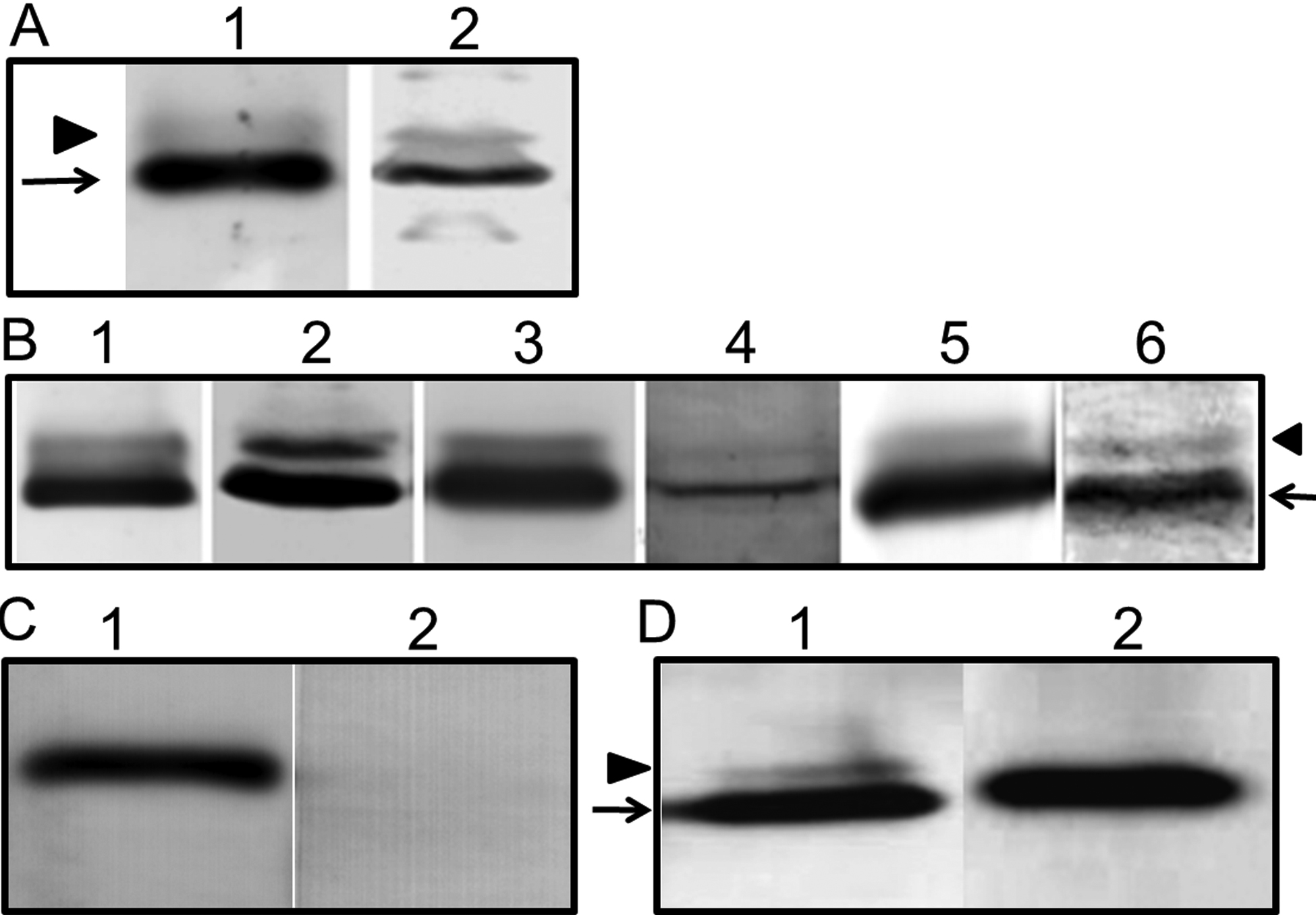
Figure 3. Immunoblot analyses of
corneal and lens membrane proteins to identify the expression of
AQP5. A: From rabbit: Lanes; 1. total cornea, 2. total
lens. B: From mouse: Lanes: 1. lacrimal gland (+ve
control), 2. total cornea, 3. total lens, 4. lens epithelial
cell membrane, 5. lens cortex fiber cell membrane, 6. lens
nuclear fiber cell membrane. C: AQP5-KO mouse lens fiber
cell membrane proteins. Lanes: 1. AQP0, 2. AQP5, expressions
studied using anti-AQP0 and anti-AQP5 antibodies, respectively.
D: Immunoblot of dephosphorylation studies in WT corneal
membrane proteins using anti-AQP5 antibody. Lanes: 1. WT
untreated proteins (arrow - ~28 kDa; arrowhead -
~34 kDa), 2. WT proteins treated with calf intestinal
alkaline phosphatase; the 34 kDa band disappeared,
presumably due to dephosphorylation.

 Figure 3
of Kumari, Mol Vis 2012; 18:957-967.
  Figure 3
of Kumari, Mol Vis 2012; 18:957-967.