Figure 2. Reverse-transcription
polymerase chain reaction (RT–PCR) analysis of AQP5. A:
In mouse lens. Lanes: 1 Wild type (WT) lacrimal gland (positive
control), 2. AQP5 knockout (AQP5-KO) lacrimal gland, 3. WT
cornea, 4. WT cornea + RNase, 5. AQP5-KO cornea, 6. WT lens
epithelium, 7. AQP5-KO lens epithelium, 8. WT lens epithelium +
RNase, 9. WT lens cortex, 10. WT lens cortex + RNase, 11.
AQP5-KO lens cortex, MW-Molecular weight marker. B:
RT–PCR analysis of AQP1 in mouse lens epithelial cells. Lanes:
1. WT lens epithelium, 2.WT lens epithelium + RNase, 3. AQP5-KO
lens epithelium, 4. AQP5-KO lens epithelium + RNase,
MW-Molecular weight marker. C: RT–PCR analysis of AQP0
in mouse lens fiber cells. Lanes: 1. WT lens cortex, 2. WT lens
cortex + RNase, 3. AQP5-KO lens cortex, 4. AQP5-KO lens cortex +
RNase, MW-molecular weight marker.
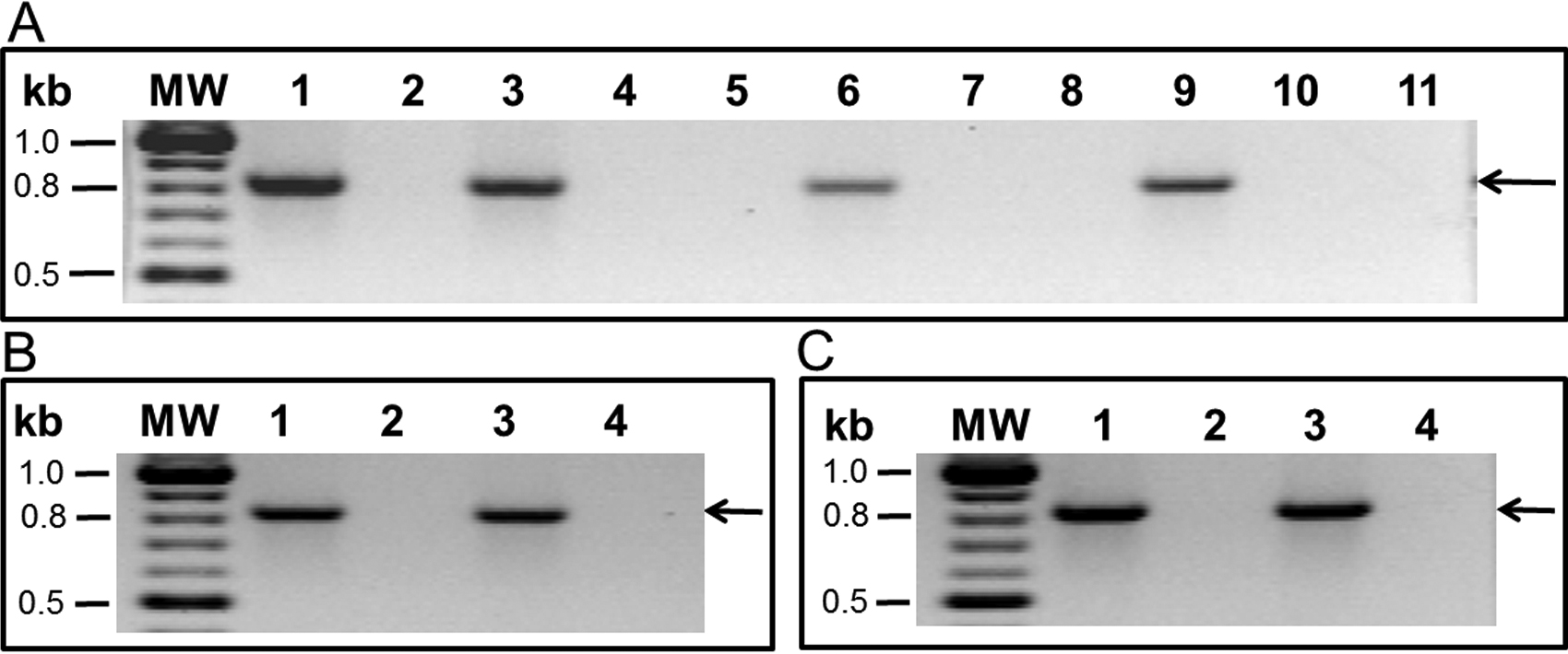
 Figure 2
of Kumari, Mol Vis 2012; 18:957-967.
Figure 2
of Kumari, Mol Vis 2012; 18:957-967.