Figure 1. Pretreatment with
intravitreal bevacizumab (IVB) significantly reduced the numbers
of vascular endothelial cells in neovascular membranes (NVMs) of
the eyes with proliferative diabetic retinopathy (PDR).
Hematoxylin and eosin stain (H&E; A, C,
and E) and von
Willebrand stain (B, D, and F) were applied to detect
the vascular endothelial cells in the NVMs of PDR eyes (A-D)
and epiretinal membranes of the eyes with proliferative
vitreoretinopathy (PVR; E, F). The untreated
group (A, B) shows a significantly more number of
vascular endothelial cells when compared to the IVB pretreated
group (C, D; p=0.003). The epiretinal membranes
of PVR eyes were set as the control group. von Willebrand stain
for vascular endothelial cells in the control group was negative
(F). Figures were selected as representative data from
three independent experiments. Scale bars: 200 μm.
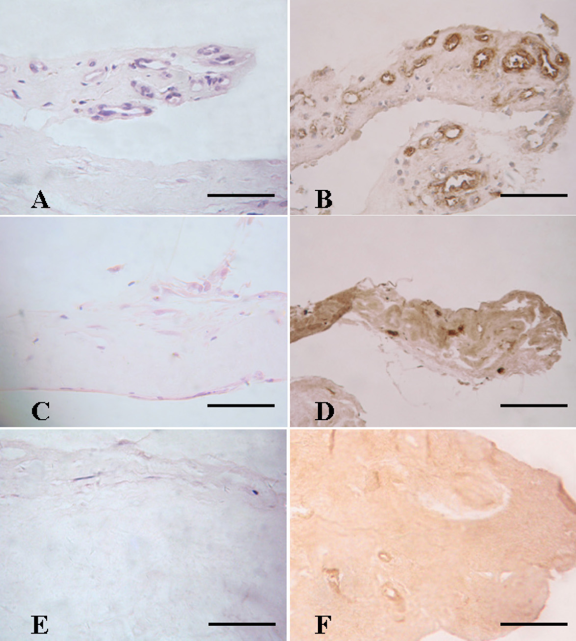
 Figure 1
of Han, Mol Vis 2012; 18:1-9.
Figure 1
of Han, Mol Vis 2012; 18:1-9.