Figure 4. Blue light damage induced tortuous photoreceptor outer segments with disrupted lamellar structures after 24 h of blue light
exposure. Transmission electron microscopy images of photoreceptors from untreated (A, C) and irradiated eyes (B, D) are presented. In part A, an overview of precisely stacked photoreceptor outer segments and the adjacent retinal pigment epithelium in the upper right
part of the image is demonstrated. In part B, an overview of light-damaged photoreceptor outer segments and their disrupted lamellar structures is presented. C: The enveloping membrane of the outer segment continuously enclosed the disks in the control retina. D: The enveloping membrane was frequently interrupted or completely lost in the middle section of the outer segment, and the
disks started to shift. Scale bars are 1 µm in A and B, and 500 nm in C and D.
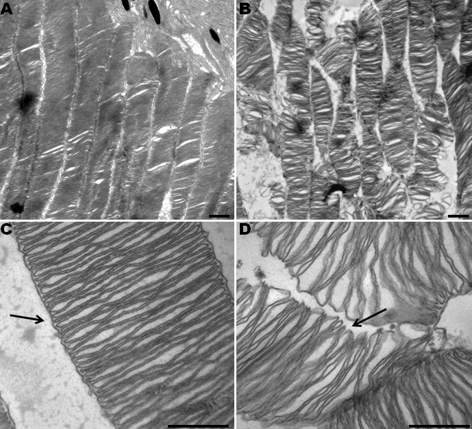
 Figure 4 of
Roehlecke, Mol Vis 2011; 17:876-884.
Figure 4 of
Roehlecke, Mol Vis 2011; 17:876-884.