Figure 1. Production of intracellular reactive oxygen species is increased by blue light damage. In part A, representative images of reactive oxygen species production by live imaging fluorescence microscopy in photoreceptor cells
of retinal explants are presented. After 1 h of blue light exposure, irradiated explants and respective non-irradiated explants
(controls) were loaded with 10 µM dihydroethidium. The scale bar represents 50 µm. In part B, quantitative analysis of reactive oxygen species production in photoreceptor cells of retinal explants is demonstrated.
Retinas were exposed to visible blue light for 0.5 h, 1 h, and 3 h. The graph displays the mean fluorescence intensity ratios
of irradiated photoreceptor cell layers versus non-irradiated time-matched controls, which are normalized to 1. Bars represent
the mean±standard error of mean (SEM) from n=5 independent experiments (*p<0.05).
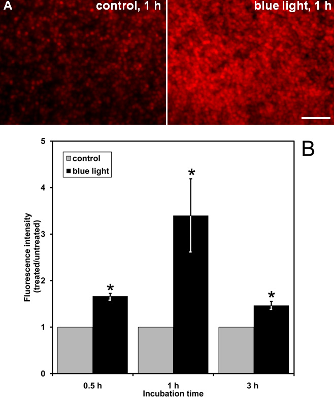
 Figure 1 of
Roehlecke, Mol Vis 2011; 17:876-884.
Figure 1 of
Roehlecke, Mol Vis 2011; 17:876-884.