Figure 3. Comparison of fundus photographs and spectral domain optical coherence tomography (SD-OCT) between patients with the p.A298D
and the p.P347L mutations. Fundus photographs (A, B, E, F) show typical retinitis pigmentosa (RP) features: retinal degeneration with pigmentation, atrophy of retinal pigment epithelium
(RPE), and attenuated vessels, which involved the area inside the vascular arcade. Compared with a 55-year-old patient with
the p.A298D mutation (A, B), a 44-year-old patient with the p.P347L mutation (E, F) had more severe retinal pigmentation, despite being 11 years younger. SD-OCT (C, D, G, H) revealed the degeneration of photoreceptor and RPE layers and the disruption of the inner and outer segment junction of
the photoreceptor in both patients. In particular, severe foveal atrophy in the patient with the p.P347L mutation resulted
in a large decrease of the central foveal thickness: 161 μm in the right eye (G; arrow) and 152 μm in the left eye (H; arrow).
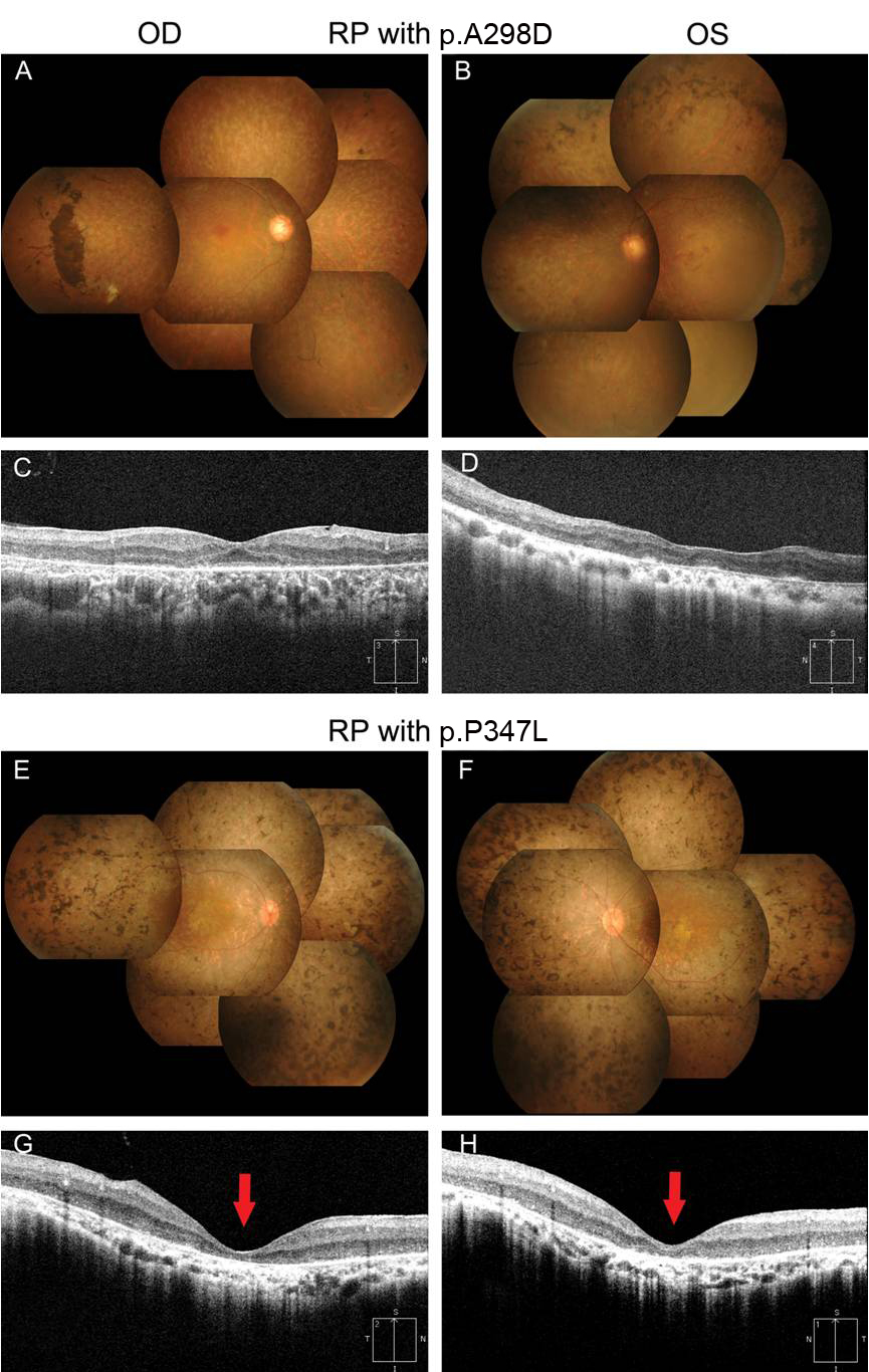
 Figure 3 of
Kim, Mol Vis 2011; 17:844-853.
Figure 3 of
Kim, Mol Vis 2011; 17:844-853.