Figure 2. Double staining immunohistochemistry was performed for TF (red) and α-SMA (green) in pterygial tissue. A-D: α-SMA immunoreactivity is colocalized with TF-positive areas in pterygial epithelial cells (D, arrows). E-H: TF immunoreactivity is detected in the other part of epithelial cells negative for α-SMA. The scale bar represents 50 μm.
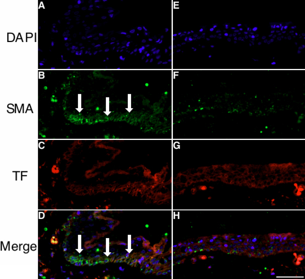
 Figure 2 of
Ando, Mol Vis 2011; 17:63-69.
Figure 2 of
Ando, Mol Vis 2011; 17:63-69.