Figure 1. Immunohistochemistry for tissue factor (TF) in a human pterygium and normal conjunctiva. Left panels are H&E staining and
right panels are TF immunoreactivity in two representative cases of a pterygium. TF is expressed in the cytoplasm of basal
(B; red arrow head), suprabasal (B; blue arrow head), and superficial cells (A; black arrow head). In the normal conjunctiva, however, immunoreactivity for TF is not detected (C). The scale bar represents 50 μm.
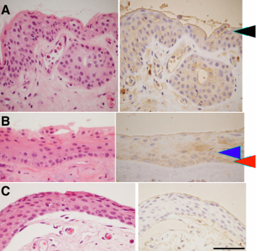
 Figure 1 of
Ando, Mol Vis 2011; 17:63-69.
Figure 1 of
Ando, Mol Vis 2011; 17:63-69.