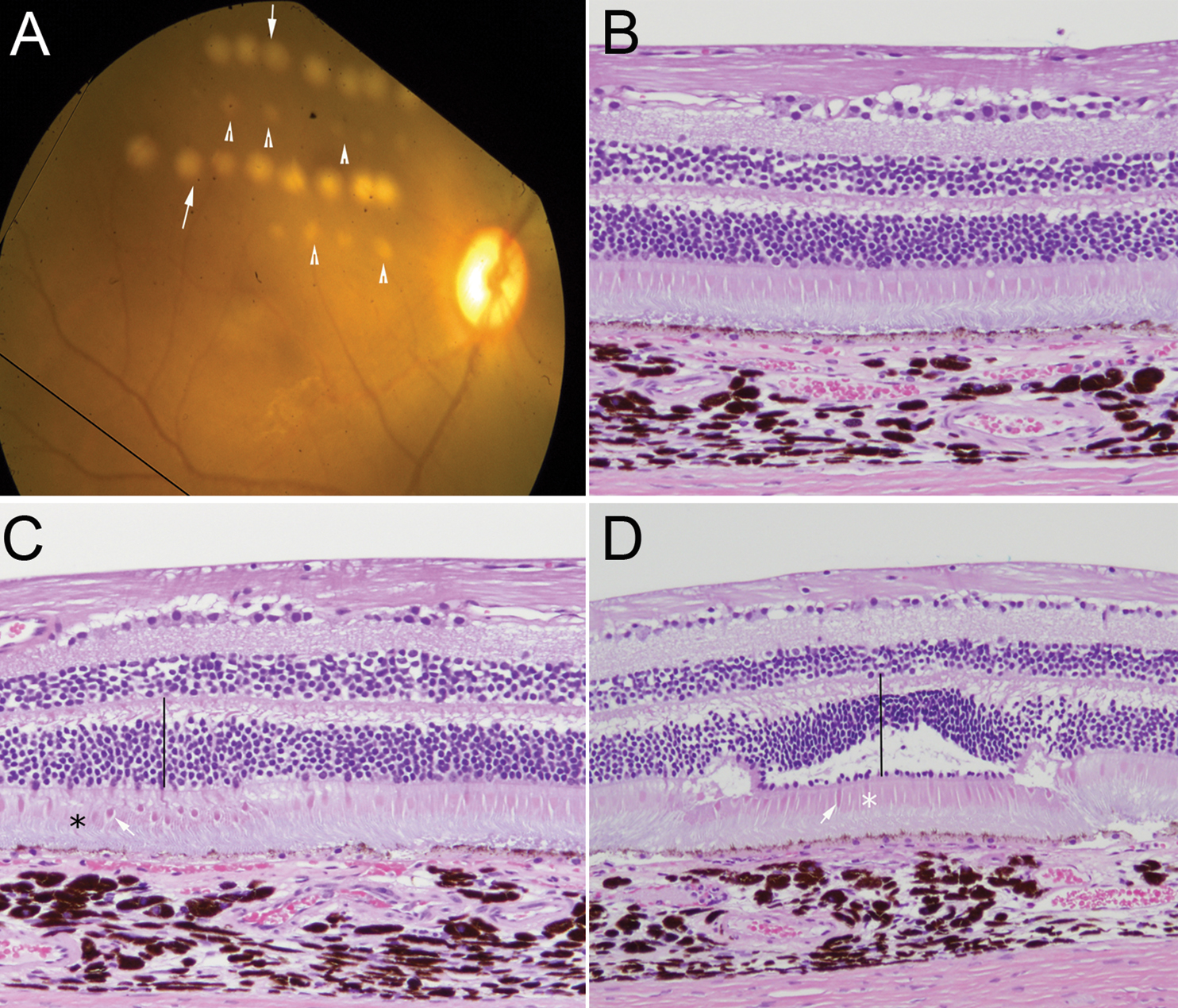
Figure 1. Fundus examination and
histological evaluation of severe and MVL type laser lesions in a
retina.
A: Rows of severe lesions (arrows) and MVL (arrow
heads) caught by fundus imaging immediately following laser treatment.
B-
D:
Histology
of normal and laser-treated retina 1 day following laser
injury.
B: Normal retina adjacent to the region that was
treated with laser. Note the intact sensory retina and retinal pigment
epithelium.
C: Minimally visible lesions show mild swelling of
the outer nuclear layer (line), condensation of cone inner segments
(arrow), and mild disruption of photoreceptor outer segments (*) and
RPE. The inner retina and underlying choroid are unaffected.
D:
A severe retinal lesion showing outer retinal swelling with disruption
of the outer nuclear layer, the outer plexiform layer, and portions of
the inner nuclear layer (line). Note the mummification of photoreceptor
inner segments (*), as indicated by shrinkage and condensation (arrow).
Also note mummification [
69,
70] of the underlying
photoreceptor outer segments and RPE (hematoxylin and eosin, original
magnification 20×).
 Figure 1 of Dunmire, Mol Vis 2011; 17:779-791.