Figure 4. Immunofluorescence for αA-crystallin and βB1-crystallin during embryonic lens development. Sections double-stained with αA-crystallin
and βB1-crystallin antisera at different developmental stages, analyzed by confocal microscopy. Some cells express predominantly
βB1-crystallin (red) and some αA-crystallin (green). Overall, there is a strong co-localization of these two crystallin proteins
throughout the lens cells. First positive immunofluorescence was detected at stage 29/30 (A). At stage 32 (B), a number of cells in the area of the lens rudiment where lens fibers will form. With further differentiation, the lens
primary fibers and secondary primary fibers are formed during stage 34–46 (C-H). Negative controls: I (without antibodies); J (only secondary antibodies); K (only primary antibodies); L: differential interference contrast (DIC). Abbreviations: Le, lens; PLF, primary lens fiber; SLF, secondary lens fiber.
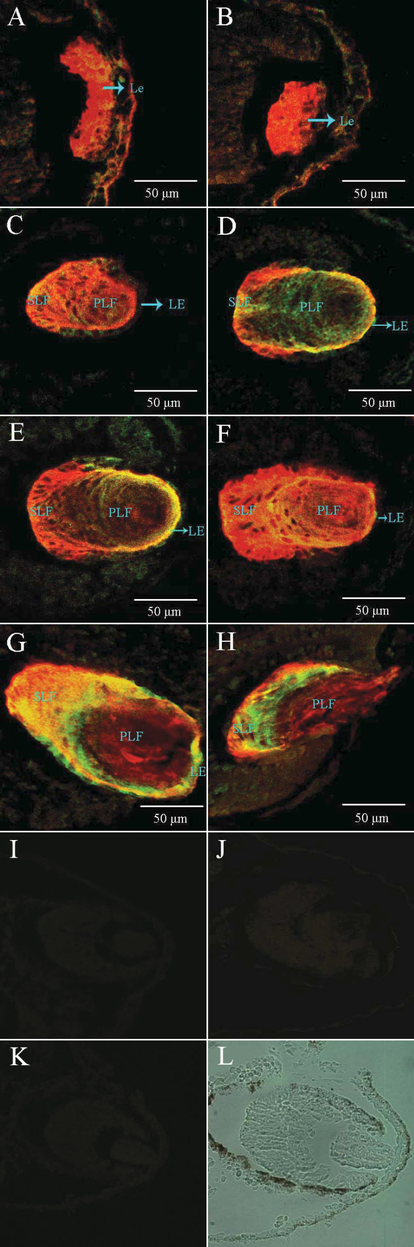
 Figure 4 of
Zhao, Mol Vis 2011; 17:768-778.
Figure 4 of
Zhao, Mol Vis 2011; 17:768-778.