Figure 3. This figure displays the optical apparatus used for human tear film thickness measurements. The laser and the camera assembly
were placed on mechanical stages equipped with X-Y-Z movement. The imaging system is mounted onto a track for scanning purposes.
Subjects placed their head on the chin-rest and maintained a constant focal length with respect to the camera assembly. Data
were streamed to a computer for further computational analysis.
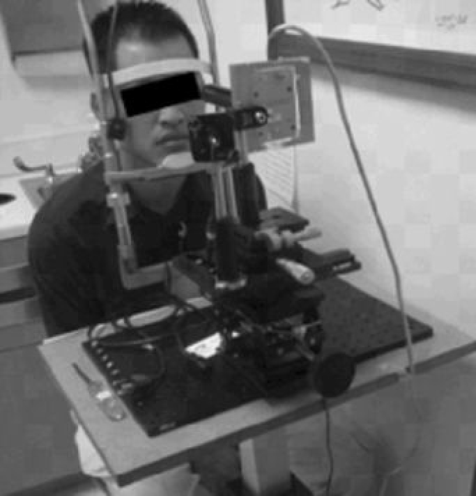
 Figure 3 of
Azartash, Mol Vis 2011; 17:756-767.
Figure 3 of
Azartash, Mol Vis 2011; 17:756-767.