Figure 4. Proliferation analysis. Immunolocalization of Ki67 positive cells (green) on 3D-HCE samples after 24h of incubation with PBS
(A), 0.01% BAC (B), KETO-BAC(-) (C), KETO-BAC(+) (D), NAA-BAC(-) (E), OPA-BAC(+) (F), CRO-BAC(-) (G), EPI-BAC(+) (H), LEVO-BAC(+) (I). Nuclei were stained with propidium iodide (PI, red). PBS (A), KETO-BAC(-) (C), NAA-BAC(-) (E) and CRO-BAC(-) (G) showed a weak expression of Ki67 in all epithelial layers. BAC at 0.01% (B), KETO-BAC(+) (D), OPA-BAC(+) (F), and EPI-BAC(+) (H) showed a higher Ki67 expression in all epithelial layers too. With LEVO-BAC(+), no Ki67 positive cells were observed, most
likely due to the deep impairment of corneal cells with a most likely inhibition of proliferative capabilities of 3D-HCE submitted
at this higher concentration in BAC. Quantification of Ki67-positive cells was concordant with these observations (J). *Statistically significant compared to PBS with p<0.04. **Statistically significant compared to 0.01% BAC with p<0.001.
$Statistically significant compared to the other solutions tested. Results are expressed as cell number per mm of epithelial
length (mm.E.L.): Mean±SD.
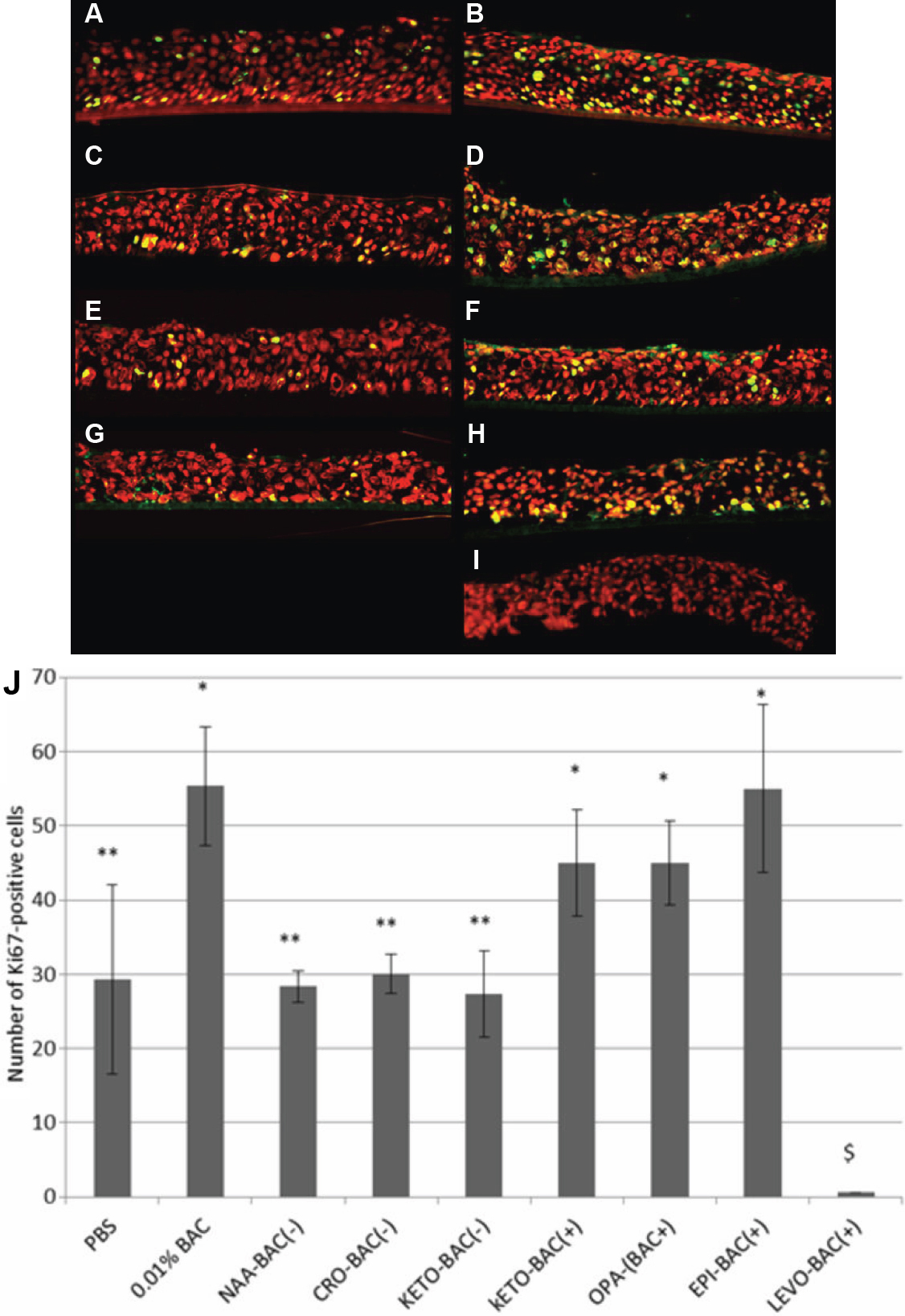
 Figure 4 of
Pauly, Mol Vis 2011; 17:745-755.
Figure 4 of
Pauly, Mol Vis 2011; 17:745-755.