Figure 6 of
Watson, Mol Vis 2011; 17:737-744.
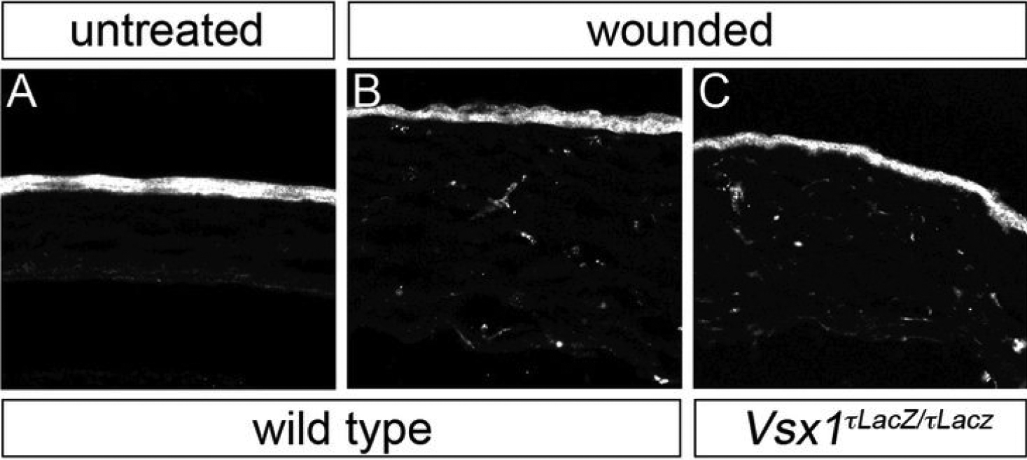
Figure 6.
Non-specific immunolabeling of alkali-wounded corneas. Panels
A
-
C
correspond to panels
I
,
M
, and
Q
, respectively, from
Figure 5
. The image pixel intensity levels have been elevated by an order of magnitude.
 Figure 6 of
Watson, Mol Vis 2011; 17:737-744.
Figure 6 of
Watson, Mol Vis 2011; 17:737-744.