Figure 2 of
Hu, Mol Vis 2011; 17:715-722.
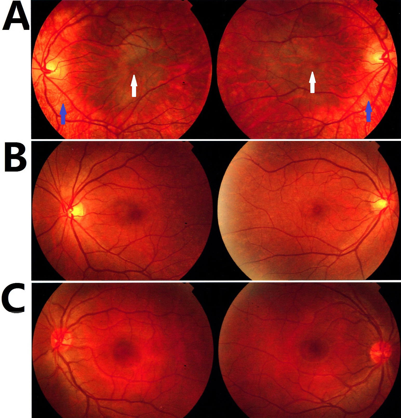
Figure 2.
Fundi photographs.
A
: Fundus of the proband (V4) revealed severe fundus hypopigmentation (blue arrow) and foveal hypoplasia (white arrow).
B
: The fundus of the carrier mother (IV9).
C
: Normal fundus (IV10).
 Figure 2 of Hu, Mol Vis 2011; 17:715-722.