Figure 5. Northern blot analysis. Northern blot of M1-M5 mRNA expression in 6 weeks minus lens treated sclera (A) and with atropine or saline treated sclera (B). Total RNA (25 µg) was loaded in each lane, run on a 1% agarose gel, transferred to a positively charged nylon membrane,
and hybridized to a fluorescein-labeled mouse M1 EcoRI enzyme digested insert cDNA clone. In the upper panel the sizes of
28S (4.7 kb), 18S (1.9 kb) rRNA and M1-M5 (2.6 kb, 1.8 kb, 3.2 kb, 1.6 kb and 3.5 kb, respectively) are indicated to the left. A: Lane 1: Mouse brain cerebellum (positive control), lane 2: minus lens treated myopic sclera, lane 3: minus lens control
sclera, lane 4: naive sclera. B: Lane 1: atropine treated myopic sclera, lane 2: atropine treated control sclera, lane 3: saline treated myopic sclera, lane
4: saline treated control sclera.
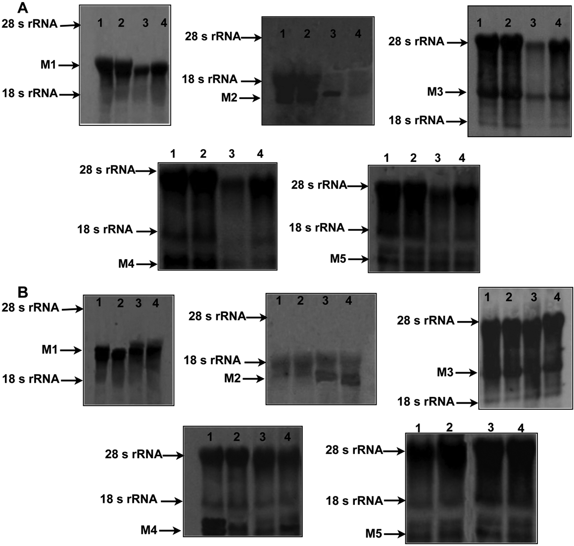
 Figure 5 of
Barathi, Mol Vis 2011; 17:680-692.
Figure 5 of
Barathi, Mol Vis 2011; 17:680-692.