Figure 3. Atropine sulfate (pan muscarinic antagonist) and normal saline treated B6 mice ocular biometry measurements were plotted against
induction period (weeks). One group was treated with sub-conjunctival injection of 1% atropine sulfate (pan muscarinic antagonist)
and another group was treated with 0.9% normal saline for four weeks. The drug treatment was started after 2 weeks of minus
lens wearing. Four weeks after drug treatment, refractive error and ocular biometry determined as before. The corneal thickness
(A) and anterior chamber depth (B) was not significantly different with or without drug treatment. The lens thickness (C), vitreous chamber depth (D) and axial length (E) were significantly reduced after receiving atropine and there was no effect with saline treatment. The myopic eye received
atropine sulfate was shifted from myopic to hyperopic when compared to saline treated myopic eye was till at myopic shift
(F). There was no significant difference seen in the control eyes. The corneal curvature was determined by automated photokeratometry.
The minus lens wearing eye’s corneal curvature was slightly flatter than the control and atropine treated eyes (G). This was significant at 4 and 6 weeks after induction of myopia. Spectacle lens-induced myopic eye received 1% atropine
for 4 weeks was significantly reduced the axial elongation, lens thickness and vitreous chamber elongation however there was
no effect with saline treatment. This result suggests that 1% atropine reduces myopia progression in mice and also no difference
in strains. These results confirm that atropine is effective in reducing myopia progression in both pigmented and non-pigmented
eyes. Data was represented as mean±S.D, *represents significance level p<0.05 and **represents significance level p<0.01.
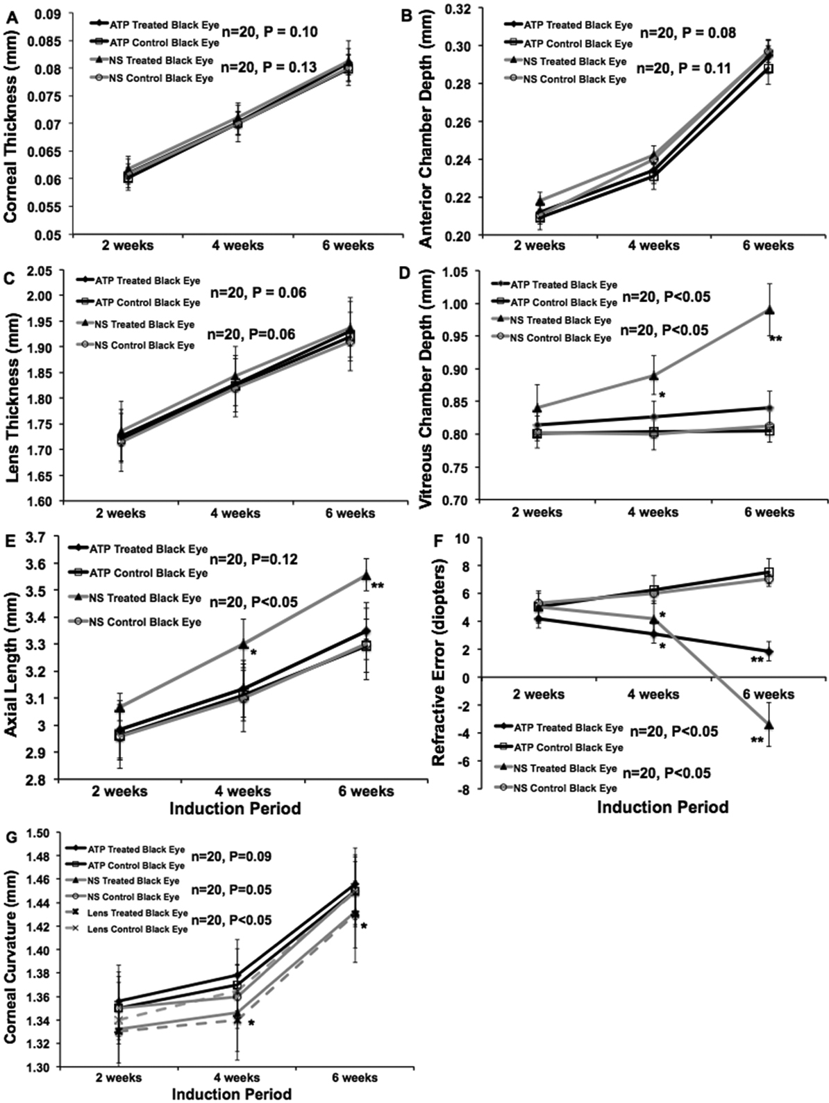
 Figure 3 of
Barathi, Mol Vis 2011; 17:680-692.
Figure 3 of
Barathi, Mol Vis 2011; 17:680-692.