Figure 5. Expression of corneal
differentiation marker in mouse corneas after limbus to limbus
epithelial scraping and treatment with conditioned media. Corneas
treated with skin conditioned media showed no K12 (A, B)
with abundant expression of K8 (C, D). Eyes treated with
limbal fibroblast conditioned media demonstrated K12 staining (E,
F) and reduced K8 positive cells (G, H).
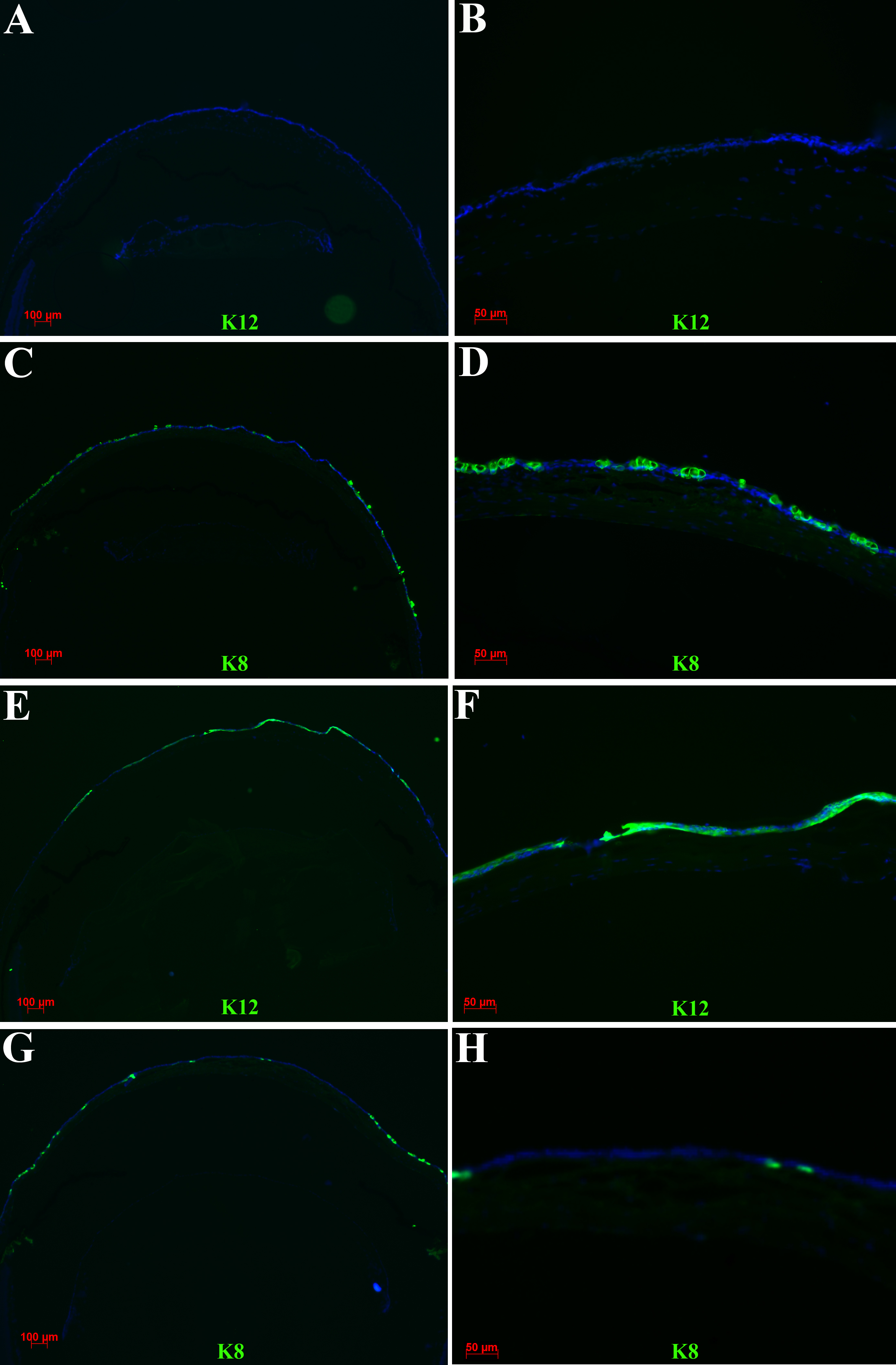
 Figure 5 of Amirjamshidi, Mol Vis 2011; 17:658-666.