Figure 4. Whole mount staining of mouse
corneas after limbus to limbus scraping followed by 3 weeks of
treatment. Corneas treated with limbal fibroblast conditioned media had
much fewer K8 positive goblet cells in the central cornea (green A,
C, D, F) but showed consistent presence of K12
positive corneal epithelial cells (red, B, C, E,
F). Corneas treated with DMEM (G-I) and skin
conditioned media (J-L) demonstrated more abundant K8 positive
cells (green, G, I, J, L) and minimal
to no K12 staining (red, H, I, K, L). A
magnified view with DAPI is shown in M and N.
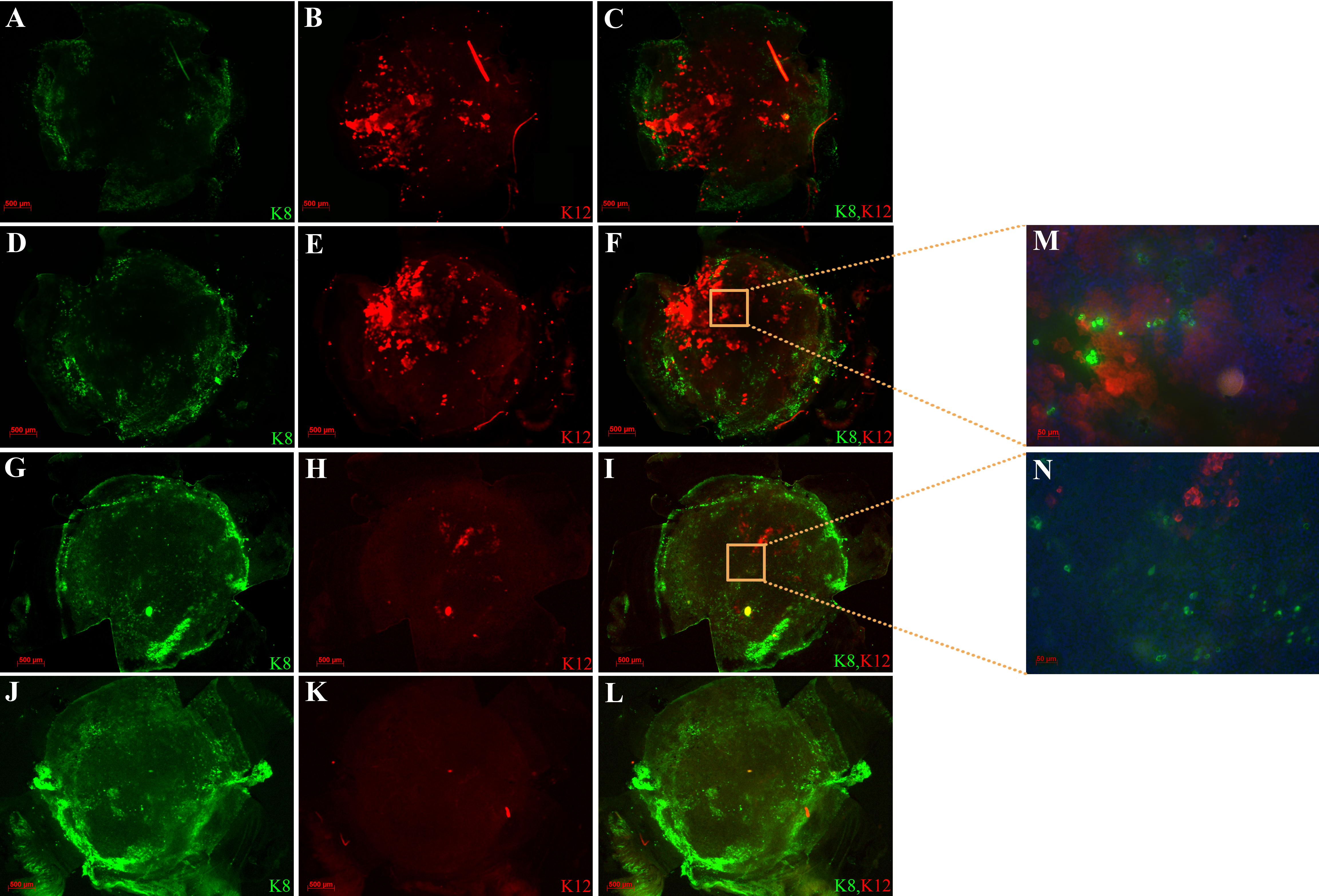
 Figure 4 of Amirjamshidi, Mol Vis 2011; 17:658-666.