Figure 3. Expression of keratin 8
(K8) and keratin 12 (K12) in normal and limbal deficient mouse corneas.
Cross sections of normal (A-D) and limbal stem cell
deficient corneas after limbus to limbus epithelial debridement (E-H).
Normal
corneal epithelium expressed K12 (A, B) but no K8
staining (C). Staining for K8 is evident only in the conjunctiva
and limbal area (D). By contrast, limbal deficient corneas
demonstrated no K12 (E, F) but abundant K8 (G, H)
staining.
Nuclei are stained with DAPI in blue.
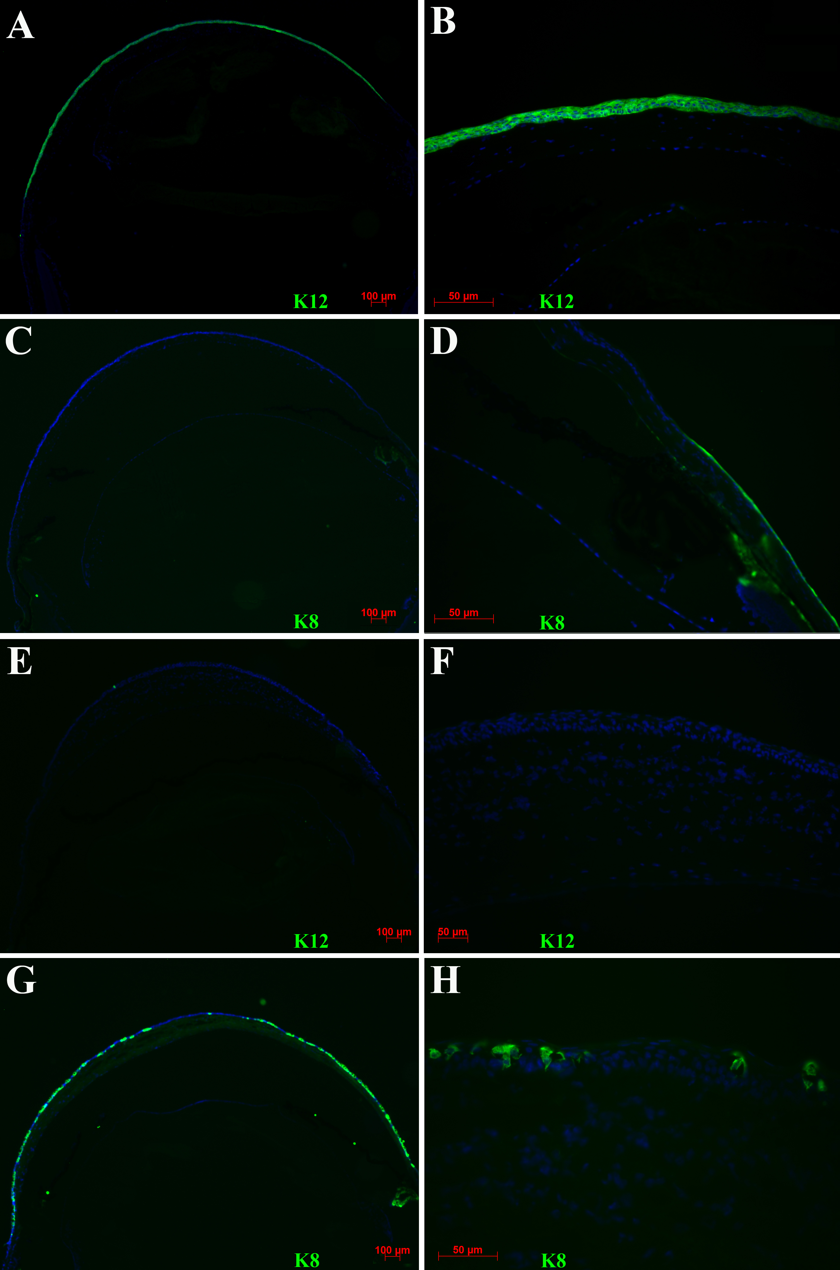
 Figure 3 of Amirjamshidi, Mol Vis 2011; 17:658-666.