Figure 2. Expression of keratin 8
(K8) and keratin 12 (K12) in normal and limbal deficient mouse corneas.
Normal (A-C) and limbal stem cell deficient corneas after
limbus to limbus epithelial debridement (D-F). Wholemount
staining shows the absence of goblet cell marker K8 (green, A),
and the presence of K12 (red, B) in a normal cornea while in
limbal deficient corneas there was the presence of K8 (green, D)
and
absence of K12 (red, E). An overlay of the K8 and K12
staining is shown for normal (C) and limbal deficient (F)
corneas. High magnification of the double staining along with DAPI
staining (blue) is shown for normal (G) and limbal deficient (H)
corneas.
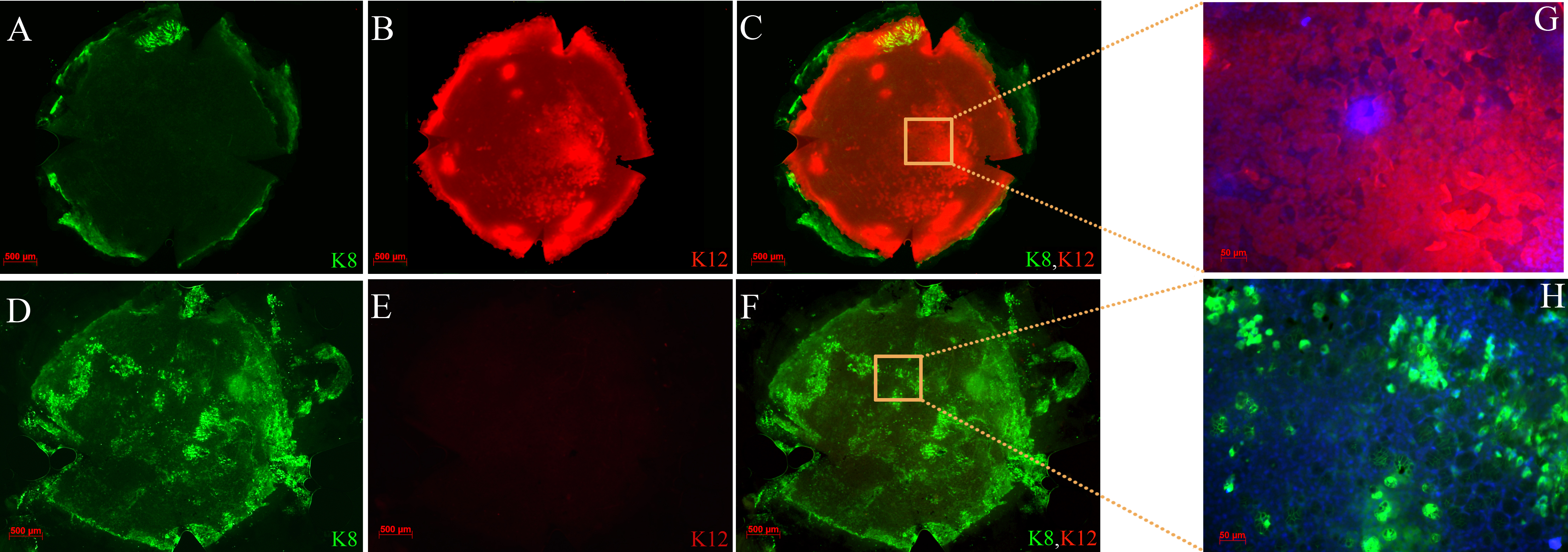
 Figure 2 of Amirjamshidi, Mol Vis 2011; 17:658-666.